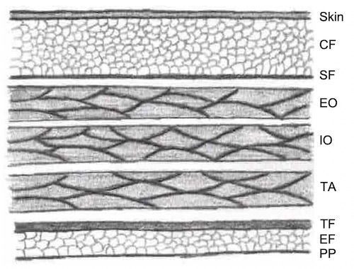
Layers of the Abdominal Wall
Above the Arcuate Line
Below the Arcuate Line

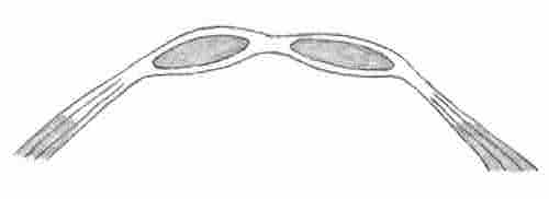
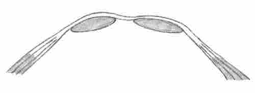
Inguinal Rings 1
Groin Ligaments
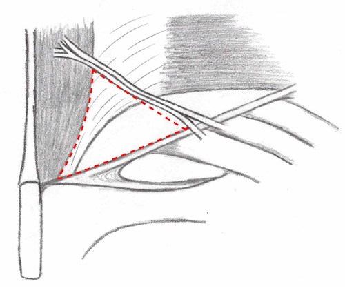
Hasselbach’s Triangle
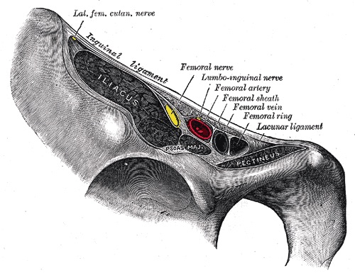
Femoral Canal 1
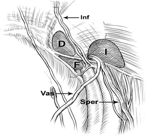
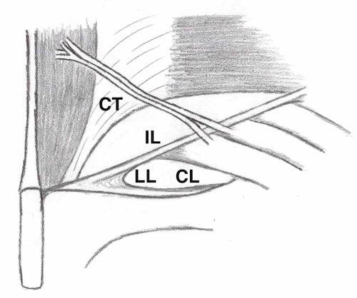
Laparoscopic Inguinal Triangles 2
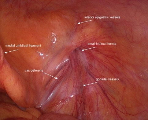
Laparoscopic Inguinal Triangles 2
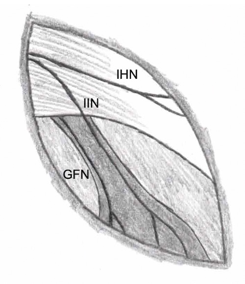
Nerves of the Inguinal Canal
Layers of the Abdominal Wall
Above the Arcuate Line
Below the Arcuate Line
Inguinal Rings 1
Groin Ligaments
Hasselbach’s Triangle
Femoral Canal 1
Laparoscopic Inguinal Triangles 2
Laparoscopic Inguinal Triangles 2
Nerves of the Inguinal Canal