Abdominal Wall: Abdominal Wall Reconstruction/Component Separation

Anterior Component Separation (ACS)

Basic Procedure

  • Isolation & Division of the External Oblique
    • Releases 7-10 cm Per Side (Total of Up to 20 cm)
  • Need More Length: Incise Posterior Rectus Sheath Above Arcuate Line
    • Adds an Additional 2-4 cm of Length

Procedure

  • 1. Subcutaneous Flaps
    • Extend to Anterior Axillary Line (Lateral of External Oblique Insertion)
    • Large Flaps Are the Largest Source of Morbidity
  • 2. Incision of External Oblique Aponeurosis
    • 2 cm Lateral to Semilunar Line/Rectus Sheath
    • Height: From Costal Margin to Pubis
  • 3. Free External Oblique from Internal Oblique
    • Extend as Far Laterally as Possible

Reconstruction

  • Consider Onlay (Subcutaneous) or Underlay (Preperitoneal) Mesh
  • Reapproximate Anterior Rectus Sheath to Midline
  • Close Skin

Modifications

  • Perforator-Preserving Technique
    • Procedure:
      • Uses Separate Inguinal Incisions (Similar to Inguinal Hernia Repair)
      • Balloon Dissector is Placed Between Internal & External Obliques and Inflated to Create the Space
      • Complete Fascial Separation Through the Inguinal Incision
    • Allows Excision of Midline Scar with Minimal Skin Flaps & Preservation of Perforators
  • Endoscopic Technique
    • Small 1 cm Incision Made Below the Costal Margin Lateral to the Rectus Abdominis
    • Space Between Internal & External Oblique Opened, Balloon Dissected & Insufflated
    • Using Laparoscope & Two Other Ports the Plane is Dissected
    • Procedure is then Repeated on the Other Side

Anterior Component Separation 1

Posterior Component Separation (PCS) & Transversus Abdominis Release (TAR)

Basic Procedure

  • Isolation & Division of the Transversus Abdominis
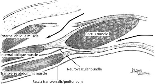
  • *Original PCS (Without TAR) Had Dissection Plane Between the Internal Oblique & Transversus Abdominis – Required Division of the Neurovascular Bundle with Denervation of Rectus Abdominis

Procedure

  • 1. Incise the Dorsal Aspect of the Posterior Rectus Sheath 1 cm from the Medial Edge of the Rectus Muscle
    • Enter the Retrorectus Space & Dissect Laterally to the Semilunar Line
    • Care to Protect the Neurovascular Bundle Laterally Near the Semilunar Line
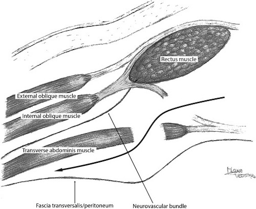
  • 2. Incise the Ventral Aspect of the Posterior Rectus Sheath at Lateral-Most Edge
    • Exposes the Underlying Transversus Abdominis
    • Do Not Penetrate Through the Transversalis Fascia/Peritoneum
  • 3. Dissect the Plane Laterally
    • Transversus Abdominis will be Anterior with the Transversalis Fascia/Peritoneum Posterior

Reconstruction

  • Reapproximate the Posterior Rectus Sheath to Midline
  • Typically Place a Sublay/Retrorectus Mesh
  • Reapproximate Anterior Rectus Sheath to Midline
  • Close Skin

Posterior Component Separation with TAR 1

Component Separation – Comparison & Complications

Comparison

  • Similar Recurrence Rate (< 10%)
  • Length Released is Debated
    • Some Report Similar Lengths
    • Many Feel ACS Allows More Medialization of the Fascia
  • PCS Mesh Extends Father Laterally – Preferred for Lateral Hernias or Ostomy
  • PCS Has Less Wound Complications – Does Not Require Large Subcutaneous Flap

Complications

  • Typically Have Improved Abdominal Wall Function
  • Surgical Site Infection
    • Most Common Complication
  • Seroma/Hematoma (2%)
  • Skin Flap Necrosis (1%)
    • Cause: Damage to Perforating Vessels
  • Iatrogenic Spigelian Hernia

References

  1. Sneiders D, de Smet GHJ, den Hartog F, Verstoep L, Menon AG, Muysoms FE, Kleinrensink GJ, Lange JF. Medialization after combined anterior and posterior component separation in giant incisional hernia surgery, an anatomical study. Surgery. 2021 Dec;170(6):1749-1757. (License: CC BY-4.0)