Anorectal: Abscess & Fistula Procedures

Anorectal/Perirectal Abscess Drainage Procedures

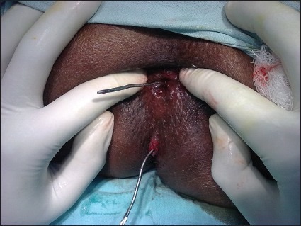
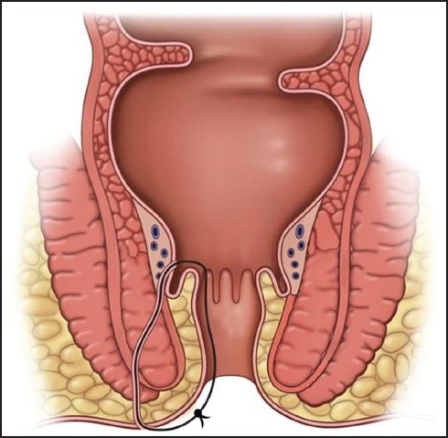
Incision and Drainage (I&D)

  • Positioning:
    • Bedside: Lateral Decubitus
    • OR: Prone Jack-Knife
  • Inject Local Anesthetic
  • Incision Over the Abscess
    • Semilunar or Cruciate (Cross)
    • Oriented Radially
    • Orient Over the Side Closest to the Anal Verge
      • Not Over the Area of Greatest Fluctuance or Area Furthest from Verge
      • Subsequent Fistula are Shorter and More Simple
    • For Large Cavities (> 5 cm): Consider Ipsilateral Counter-Incisions to Avoid an Unnecessarily Large Single Incision
  • Bluntly Probe the Cavity to Break All Loculations and Drain All Fluid Pockets
    • Avoid Over-Aggressive Disruption – Risk for Sphincter or Pudendal Nerve Injury
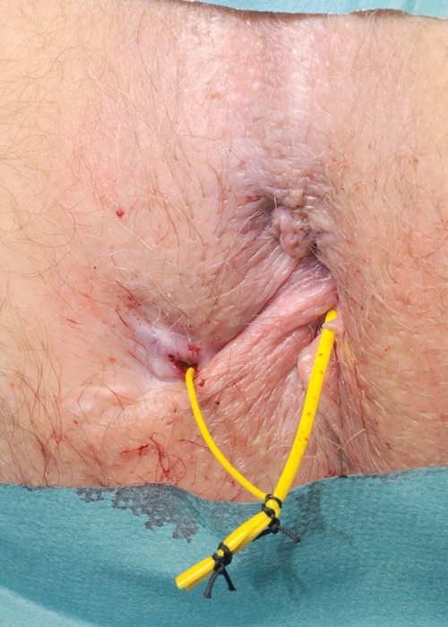
  • Finish Options:
    • May Consider Packing with Daily Changes
      • Most Common Although No High-Quality Evidence of Any Benefit
    • Excise ≥ 1 cm Segment of Skin to Prevent Premature Closure without Packing

I&D

Hanley Procedure

  • For Horseshoe Abscess
  • Traditional Hanley Procedure: 3 Incisions
    • Counter Incisions Over Each Bilateral Ileoanal Fossa
    • Deep Space: Posterior Incision Between Coccyx & Anus or Through the Internal Anal Sphincter
    • Counter Drains Placed Through Each Lateral Extension – Removed After 2-4 Weeks
    • *High Risk for Anorectal Incontinence – Divided Posterior Sphincter Mechanism
  • Modified Hanley Procedure: 2 Incisions with a Cutting Seton
    • Counter Incisions Over Each Bilateral Ileoanal Fossa
    • A Cutting Seton Over the Posterior Sphincter Mechanism is Serially Tightened Over Months
    • *Posterior Sphincter is Replaced by Tethered Scar Tissue – Decreases Risk of Incontinence

Hanley Procedure

Fistulotomy

Basics

  • Fistula is Opened to Allow Drainage and Healing by Secondary Intention

Procedure

  • Define the Fistula Tract
    • Anoscopy to Identify the Internal Opening
    • Insert a Fistula Probe Through the External Opening
    • Direct Towards the Internal Opening
      • Avoid Creating a False Passage
  • Options if Unable to Identify:
    • Inject Dilute Hydrogen Peroxide Through an Angiocatheter into the External Opening to Identify
    • Using a Curved Fistula Probe Attempt to Identify the Tract from the Internal Opening to Identify
    • Partial Fistulectomy
  • Cannulate the Fistula with a Probe
  • Divide the Tissue Overlying the Probe with Electrocautery

Outcomes

  • Success Rate: > 90%
  • Risk of Fecal Incontinence: 0-40% (Increased if Complex)

Fistula Probe 1

Setons

Basics

  • Seton Material:
    • Vessel Loop
    • Thin Silastic Band
    • Silk Suture
  • Types:
    • Noncutting/Draining Seton – Loose Connection at Skin Level
      • Allows Continued Drainage & Promotes Fibrosis/Maturation
      • Often Requires a Second-Stage Procedure Once Mature
        • Procedures: Fistulotomy, LIFT or Advancement Flap
        • Timing: After 6-10 Weeks
          • May Wait 3-4 Months if More Complex
      • May Be Definitive Treatment in Crohn’s or High-Risk Patients
    • Cutting/Snug Seton – Tight Connection at Skin Level
      • Slowly Tightened Every 4-6 Weeks
      • Must Incise Skin Surface at Initial Placement – Cutting Seton on Skin is Painful
      • Mostly Abandoned Due to High Risk of Fecal Incontinence

Placement

  • Define the Fistula Tract (Similar to Fistulotomy)
  • A Seton is Passed Through the Fistula Tract & Secured to Itself in a Loop or Omega Shape

Outcomes

  • Success Rate:
    • Noncutting: 62-100%
    • Cutting: 82-100%
  • Risk of Fecal Incontinence:
    • Noncutting: Rare
    • Cutting: 30%

Seton 2

Seton Diagram 3

Ligation of Internal Fistula Tract (LIFT Procedure)

Basics

  • Typically Preformed After Tract is Matured
  • Fistula Tract is Divided in the Intersphincteric Space

Procedure

  • Cannulate the Tract with a Fistula Probe
  • Make a Skin Incision Over the Intersphincteric Groove
  • Dissect Down the Intersphincteric Space to the Deep Fistula Tract
  • Bluntly Define & Isolate the Tract Circumferentially
  • Remove Probe
  • Suture Ligate the Intersphincteric Tract & Sharply Divide
  • Close the Skin

Outcomes

  • Success Rate: 61-95%
  • Risk of Fecal Incontinence: Rare

Endoanal/Endorectal Advancement Flap

Basics

  • Resect Mucosa Around the Internal Opening and Pull-Down Rectal/Anal Mucosa to Cover
  • Dermal Advancement Flap
    • Pulls Up Skin Instead
    • Preferred if Below Dentate Line to Prevent Mucosal Ectropion with Pruritis & Mucosal Drainage

Procedure

  • Create a Flap of Mucosal Tissue Proximal to the Internal Opening
    • Include Mucosa Around the Internal Opening
    • Must Be Wide-Based (Width ≥ 2-3x Length) – Ensure Good Perfusion
    • Excise Flap Tip Containing the Internal Mucosal Opening
  • Manage the Tract
    • Debride the Tract with a Curette
    • Close Internal Opening with Suture
    • Widen the External Opening to Ensure Good Drainage
  • Pull Flap Distally and Reapproximate the Mucosal Edges

Outcomes

  • Success Rate: 60-100%
  • Risk of Fecal Incontinence: 0-12.5%

References

  1. Dutta G, Bain J, Ray AK, Dey S, Das N, Das B. Comparing Ksharasutra (Ayurvedic Seton) and open fistulotomy in the management of fistula-in-ano. J Nat Sci Biol Med. 2015 Jul-Dec;6(2):406-10. (License: CC BY-NC-SA-3.0)
  2. Tanner NC, Maw A. A novel technique for negotiation of a complex fistula-in-ano using a flexible ureteral catheter. Ann R Coll Surg Engl. 2014 Jan;96(1):80. (License: CC BY-3.0)
  3. Kolar B, Speranza J, Bhatt S, Dogra V. Crohn’s disease: Multimodality Imaging of Surgical Indications, Operative Procedures, and Complications. J Clin Imaging Sci. 2011;1:37. (License: CC BY-NC-SA-3.0)