Esophagus: Barrett’s Esophagus

Barrett’s Esophagus

Basics

  • Definition: Normal Squamous Epithelium of Distal Esophagus Replaced by Metaplastic Columnar Epithelium with Goblet Cells
  • Cause: Chronic Exposure to Gastric Acid from GERD
    • Risk of Development in GERD: 6-15%
  • 30-125x Increased Risk for Adenocarcinoma

Risk Factors

  • Male
  • Age > 50
  • Caucasian
  • Obese
  • Tobacco
  • Family History

Diagnosis

  • Screening Indication: Male with Chronic (> 5 Years) or Frequent (≥ Weekly) GERD Symptoms with ≥ 2 Other Risk Factors
  • Seattle Protocol for Endoscopic Evaluation: Need 4 Quadrant Bx Every 1-2 cm of Metaplasia
    • Recommend Review by Two Separate Pathologists (High Variability Between Observers)

Prague Criteria

  • Grading Criteria Used When Reporting Extent of Disease (i.e. C5M7)
  • C Value: Circumferential Extent
    • Endoscope Depth at GE Junction Minus Depth at the Proximal-Most Circumferential Extent
  • M Value: Maximum Extent
    • Endoscope Depth at GE Junction Minus Depth at the Proximal-Most Maximum Extent

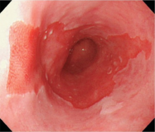
Barrett’s Esophagus 1

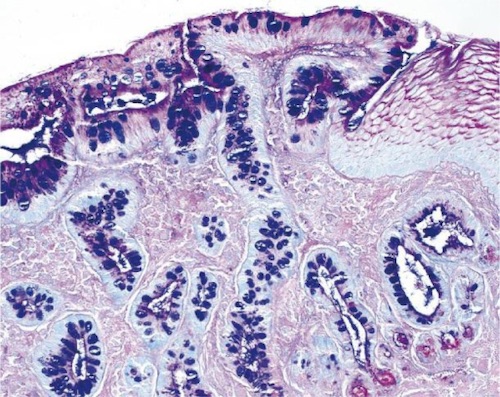
Columnar Metaplasia 2

Barrett’s Esophagus – Management

Surveillance Endoscopy Schedule

  • No Dysplasia: Every 3-5 Years
  • Indefinite for Dysplasia: Confirm At 3-6 Months, Then Every 12 Months
  • Low-Grade Dysplasia: Every 6-12 Months
  • High-Grade Dysplasia (If No Intervention): Every 3 Months

Indefinite for Dysplasia

  • Primary Treatment: PPI & Surveillance Endoscopy

No Dysplasia

  • Primary Treatment: PPI & Surveillance Endoscopy
  • If Fails: Fundoplication
  • Goal is to Prevent Symptoms & Further Metaplasia
    • CA Risk Not Proven to Be Reversible

Low-Grade Dysplasia

  • Primary Treatment: PPI & Surveillance Endoscopy
  • If Persists: Fundoplication
  • If Still Persists: Radiofrequency Ablation (RFA)

High-Grade Dysplasia

  • Primary Treatment: Endoscopic Mucosal Resection (Most Common) Esophagectomy
  • Indications for Esophagectomy:
    • Patient Preference or Unable to Comply with Endoscopic Surveillance
    • Large Lesion (> 2-3 cm)
    • Multifocal
    • Progression or Failed Ablation
    • Impaired Esophageal Function

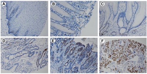
Immunohistochemical Analysis of Ki67 Expression: (A) Squamous Mucosa, (B) Columnar Metaplasia, (C) Barrett’s, (D) Low-Grade Dysplasia, (E) High-Grade Dysplasia, (F) Adenocarcinoma 3

References

  1. Japan Esophageal Society. Japanese Classification of Esophageal Cancer, 11th Edition: part I. Esophagus. 2017;14(1):1-36.(License: CC BY-4.0)
  2. Migaczewski M, Pędziwiatr M, Matłok M, Budzyński A. Laparoscopic Nissen fundoplication in the treatment of Barrett’s esophagus – 10 years of experience. Wideochir Inne Tech Maloinwazyjne. 2013 Jun;8(2):139-45. (License: CC BY-NC-ND-3.0)
  3. Choy B, LaLonde A, Que J, Wu T, Zhou Z. MCM4 and MCM7, potential novel proliferation markers, significantly correlated with Ki-67, Bmi1, and cyclin E expression in esophageal adenocarcinoma, squamous cell carcinoma, and precancerous lesions. Hum Pathol. 2016 Nov;57:126-135.(License: CC BY-2.0)