Small Intestine: Bowel Resection & Anastomosis

Bowel Resection

Principles

  • Ensure Good Blood Supply – Judge Subjectively or Use Doppler/Scintigraphy
  • Ensure Adequate Mobilization & Avoid Tension
  • Angle Transection Line Straight or Somewhat Oblique to Keep Antimesenteric Edge Shorter & Ensure Good Blood Supply

Technique

  • Classic Technique
    • Place Non-Crushing Bowel Clamps on Both Sides of the Transection Line
    • Sharply Transect the Bowel Using a Scalpel
    • Repeat at the Opposite Bowel Margin
    • Transect the Mesentery Using an Electrosurgical Device or by Clamping & Tying
  • Linear Stapler Technique (More Common)
    • Pinch & Thin the Mesentery Just Under the Site of Transection
    • Make a Small Mesenteric Defect at the Site
    • Insert One Jaw of the Stapler Through the Defect
    • Assemble & Fire the Stapler at the Transection Line
    • Repeat at the Opposite Bowel Margin
    • Transect the Mesentery Using an Electrosurgical Device or by Clamping & Tying

Bowel Anastomosis

General Principles

  • Strength Layer of Bowel: Submucosa
    • All Layers Help
    • Strongest – Esophagus Mucosa Squamous Cell Layer
  • Center Side Reconstructions On:
    • Small Bowel: Antimesenteric Border (High Risk for Ischemia if Elsewhere)
    • Large Bowel: Taenia Coli (Adds Strength)
  • Anastomosis Weakest Time Point: 3-5 Days (Breakdown Exceeds Production)
    • Increased Collagenase Compared to Skin

Type of Anastomosis

  • End-to-End
    • The End of One Loop is Anastomosed to the End of Another Loop
    • More Physiologic in Replication of Normal Gut Motility
  • End-to-Side
    • The End of One Loop is Anastomosed to the Side of Another Loop
    • Considered When There is a Size Mismatch (Small Bowel to Colon) Although Others are Still Possible
  • Side-to-Side
    • Bowel Loops are Oriented Side-to-Side in an Overlapping Fashion
    • Most Common when Using a Linear Stapler
    • Types:
      • Isoperistaltic: Ends are Approximated to Maintain Similar Directions of Peristalsis
        • Technically More Difficult
      • Antiperistaltic: Ends are Approximated with Opposite Directions of Peristalsis
        • Technically Less Difficult
        • Often Referred to as “Functional End-to-End”

Method

  • Hand-Sewn
    • Excise Staple Lines if Present
    • Use a Stay Suture to Approximate Both Bowel Ends
    • Create Anastomosis with Full-Thickness Bites Using Absorbable Suture – Techniques are Varied (Interrupted vs Running/Direction of Travel)
    • Consider Seromuscular Lembert Sutures to Buttress as a Second Layer
    • Close the Mesenteric Defect
  • Stapled
    • Traditional Method:
      • Resect Proximal & Distal Ends
      • Sharply Excise the Antimesenteric Corner of the Resected Ends
      • Place the Two Jaws of the Linear Stapler Through Each Opening
      • Arrange the Jaws Along the Antimesenteric Border
      • Assemble & Fire the Stapler to Create a Common Channel
      • Consider Placing a Reinforcing Silk Stitch (“Crotch Stich”) at the Inner Junction – The Site of Most Tension
      • Close the Enterotomies – Either with Suture or With Another Staple Load
      • Close the Mesenteric Defect
    • Barcelona Technique (For Ileostomy Reversal):
      • Approximate Proximal & Distal Ends (Prior to Resection)
      • Make Small Enterotomies at the Ends
      • Pass Stapler into Both Ends to Create Common Channel
      • Use Another Stapler to Amputate the Specimen
      • *Benefits: Cost-Effective & Only Uses Two Staple Loads (Traditional Uses 4 Staple Loads)

Comparison

  • No Significant Difference in Outcomes Between Techniques
  • No Difference Between Single-Layer vs Double-Layer Hand-Sewn Anastomoses
  • No Difference Between Hand-Sewn & Stapled Anastomoses
    • Possibly Higher Incidence of Radiographic Leaks When Hand-Sewn (12.2% vs 4.1%) But Not Clinical Leaks (3.2% vs 4.7%)
    • Conflicting Data on Ileocolic Anastomoses (Specifically)

Intraoperative Assessment of Anastomosis Viability

  • Colorectal Anastomosis Can be Endoscopically Evaluated by an Air-Leak Test
  • Assessment of Perfusion;
    • Historical Subjective Findings:
      • Bowel Color
      • Observed Pulsatile Flow at the Cut Section
    • Modern Objective Findings:
      • Indocyanine Green Fluorescence Angiography (ICG-FA/ICGA) – Best Studied
      • Doppler US – Minimal Data
      • Light Spectroscopy – Minimal Data

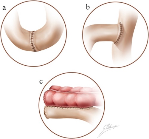
Anastomosis; (a) End-to-End, (b) End-to-Side, (c) Side-to-Side 1

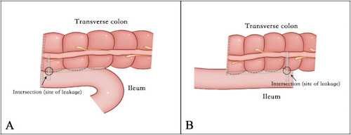
Side-to-Side Anastomosis; (A) Antiperistaltic, (B) Antiperistaltic 2

Indocyanine Green Fluorescence Angiography (ICG-FA/ICGA)

  • Indocyanine Green (ICG) is a Fluorescent Probe in Response to Near-Infrared (NIR) Light
    • Binds Primarily to Serum Albumin & Other Plasma Proteins
    • Primarily Confined to the Intravascular Compartment with Minimal Leakage
    • Excreted Almost Exclusively into Bile
    • Negligible Toxicity
  • Timing:
    • Bowel Should Enhance < 60 Seconds
    • Half-Life: 3-5 Minutes
    • Cleared by Liver: 15-20 Minutes
  • Dosing: Poorly Standardized
    • 2-0.5 mg/kg
    • 5-12.5 mg
  • Imaging Systems:
    • Firefly (Intuitive Surgical)
    • SPY Elite (Stryker)
    • PINPOINT (Stryker)
    • IMAGE1 S (Karl Storz)
    • D-LIGHT P SCB (Karl Storz)
  • Effects on Colorectal Anastomoses: Possibly Decreased Risk of Anastomotic Leak, Reoperation & Overall Complications (Debated)
    • Insufficient Data Evaluating Small Bowel Anastomoses

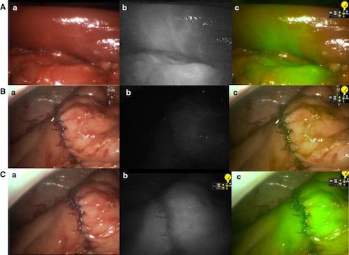
Right Hemicolectomy by ICG-FA; (A) Bowel After Vessel Division with Demarcation, (B) Anastomosis Before ICG, (C) Anastomosis After ICG; (a) Normal Light, (b) NIR, (c) Superimposition of NIR in Green 3

Anastomotic Leak

Leak Rates

  • Overall: 2-7%
  • Ileocolic: 1-3% (Lowest)
  • Colocolic: 6-12%
  • Coloanal: 10-20% (Highest)

Risk Factors

  • Patient Factors:
    • Male Gender
    • Malnutrition
    • High ASA Score
  • Emergency Surgery (Compared to Elective)
  • Operative Factors:
    • Ischemia/Tension
    • Tumor Size > 5 cm
    • Multiple Stapler Firings
    • Low Anastomosis (< 5 cm from Anal Verge)
    • Lateral LN Dissection
    • Prolonger OR (> 4 Hours)
  • Debated Risk Factors:
    • NSAIDs/Ketorolac (Toradol)
    • Corticosteroids
    • Drains

Presentation

  • Abdominal Pain
  • Fever
  • Tachycardia
  • Purulent or Feculent Drainage
  • May present with an Abscess/Fluid Collection

Treatment

  • Subclinical/Radiographic: Conservative Management
  • Small (< 3 cm) Contained Abscess: Conservative Management & ABX
  • Large (> 3 cm) or Multiloculated Abscess: Percutaneous Drain
    • If Fails: Surgical Drainage
  • Unstable, Peritonitis or Free Intraperitoneal Leak: Surgical Repair

References

  1. Terrone DG, Lepanto L, Billiard JS, Olivié D, Murphy-Lavallée J, Vandenbroucke F, Tang A. A primer to common major gastrointestinal post-surgical anatomy on CT-a pictorial review. Insights Imaging. 2011 Dec;2(6):631-638. (License: CC BY-2.0)
  2. Zhang M, Lu Z, Hu X, Zhou H, Zheng Z, Liu Z, Wang X. Comparison of the short-term outcomes between intracorporeal isoperistaltic and antiperistaltic totally stapled side-to-side anastomosis for right colectomy: A retrospective study on 214 consecutive patients. Surg Open Sci. 2022 Mar 26;9:7-12. (License: CC BY-NC-ND-4.0)
  3. Ris F, Hompes R, Cunningham C, Lindsey I, Guy R, Jones O, George B, Cahill RA, Mortensen NJ. Near-infrared (NIR) perfusion angiography in minimally invasive colorectal surgery. Surg Endosc. 2014 Jul;28(7):2221-6. (License: CC BY-4.0)