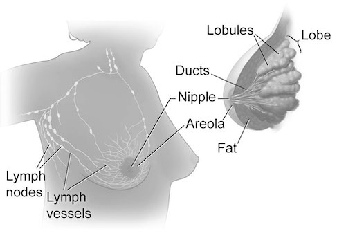
Breast Anatomy 1
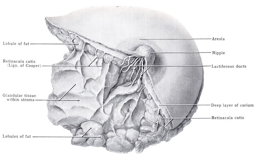
Cooper’s Ligaments 2
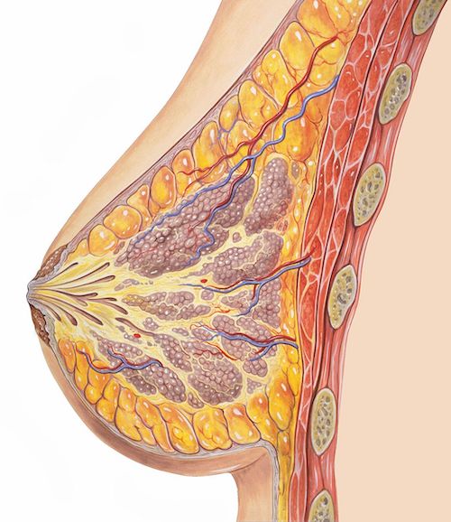
Breast Cross-Section 3
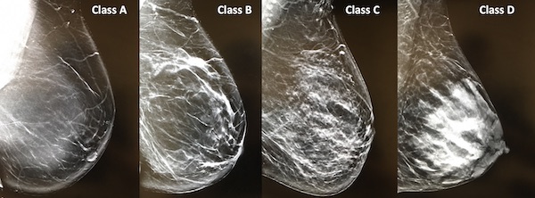
Breast Density
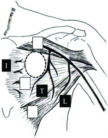
Breast Blood Supply: Internal Mammary Artery (I), Thoracoacromial Artery (T), Lateral Thoracic Artery (L) 4
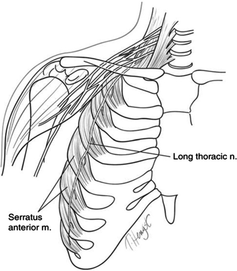
Long Thoracic Nerve 5
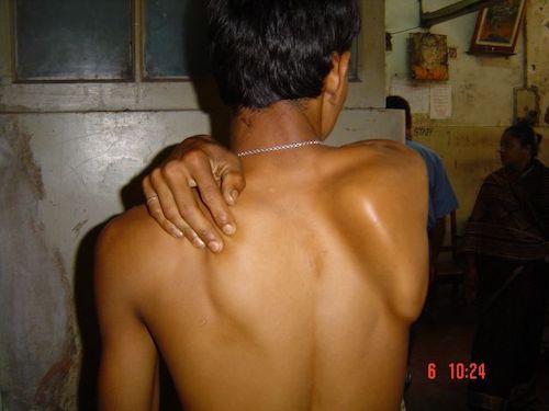
Winged Scapula 6
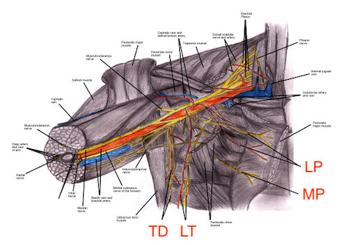
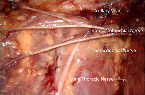
Axillary Nerves 7
Intercostal Brachial Nerve 8
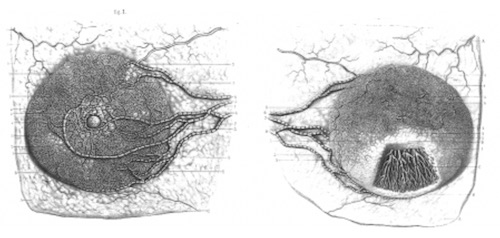
Sappey Plexus 9
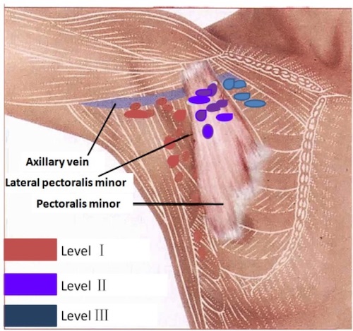
Axillary Lymph Nodes Levels 10
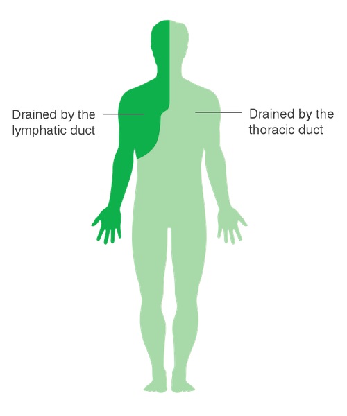
Body Lymphatic Drainage 11