Small Intestine: Carcinoid Tumor

Carcinoid Tumor

Pathophysiology

  • Well-Differentiated Neuroendocrine Tumor Arising from Gastrointestinal Enterochromaffin (Kulchitsky) Cells
  • Produce:
    • Serotonin (Metabolized to 5-HIAA)
    • Kallikrein
    • Histamine
    • Tachykinins
    • Prostaglandin

Location

  • Most Common Site: Small Intestine (50%) < Rectum < Appendix < Colon < Stomach
    • Most Common in Ileum
    • *Past Believed That the Appendix Was Most Common
  • Most Common Primary Small Intestine Tumor
  • Gastric Tumor Types (If in Stomach)
    • Type I: Associated with Chronic Atrophic Gastritis & Pernicious Anemia
      • Most Common Gastric Subtype (70-80%)
      • Grow Slowly
    • Type II: Associated with Gastrinoma & Possibly MEN-I
      • Intermediate Growth
    • Type III: Sporadic
      • Aggressive

Presentation

  • Most Common Presenting Sx: Abdominal Pain
  • Carcinoid Syndrome
    • Infrequently Encountered
    • Occurs Once Tumor Metastasizes to Liver or Retroperitoneum
      • Serotonin Cleared in Liver & Lung by Monoamine Oxidase if Only Produced by GI Tract
    • Sx:
      • Flushing (From Kallikrein)
        • Clinical Hallmark of Carcinoid Syndrome (85%)
      • Diarrhea (From Serotonin)
      • Bronchoconstriction (From Bradykinin)
      • Right Heart Valve Lesions & Tricuspid Insufficiency
      • Venous Telangiectasia (From Prolonged Vasodilation of Face)
  • Carcinoid Crisis
    • The Release of an Overwhelming Amount of Hormone from the Tumor
    • Causes Hemodynamic Instability & Symptoms of Carcinoid Syndrome
    • Triggers:
      • Anesthesia Induction
      • Tumor Manipulation (Biopsy or Manual Palpation)
      • Invasive Procedures
      • Less Common:
        • Chemotherapy
        • Hepatic Artery Embolization

Prognosis

  • 25% Have More Than One Small Bowel Tumor at Diagnosis
  • 40-80% Have Spread to the Mesentery with Nodal Metastases at Diagnosis
  • 2/3 Have Spread by Time of Dx (Local or Systemic)

Diagnosis

  • CT of Mesenteric Masses Classically Show a Spiculated Mass with Calcification & Desmoplastic Reaction
  • Detect:
    • Urinary 5-HIAA
      • Best Initial Test
      • Low Sensitivity if Non-Functioning
    • Chromogranin A Level
      • Can Also Detect Non-Functioning
      • Low Specificity (Not for Diagnosis)
      • Use: Progression, Response & Recurrence
  • Localize:
    • Octreotide Scan
    • 68 Ga-DOTATATE PET/CT Scan – Generally Preferred if Available
      • Superior Image Quality with Greater Patient Convenience (Octreotide Scan May Require Repeat Images the Next Day)

Treatment

  • Primary Tx: Resection
    • If Unresectable: Chemo (Streptozocin & 5FU) & Octreotide
  • Liver Mets: Resect if Able
    • Contraindications:
      • Diffuse Bilobar Involvement
      • Compromised Liver Function
      • Extrahepatic Metastases
      • Presence of Poorly-Differentiated Carcinoma
    • Consider Preoperative Hepatic Artery Embolization
    • Very Good Survival Rates Even with Extensive Liver Mets
  • Symptomatic Tx:
    • Flushing: α-Blockers
    • Other Symptoms: Octreotide
  • Carcinoid Crisis: Octreotide

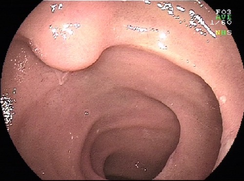
Carcinoid Tumor in Ileum 1

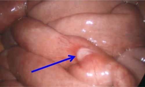
Carcinoid Tumor in Jejunum 2

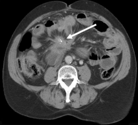
Carcinoid Tumor with Calcified Mesenteric Mass 3

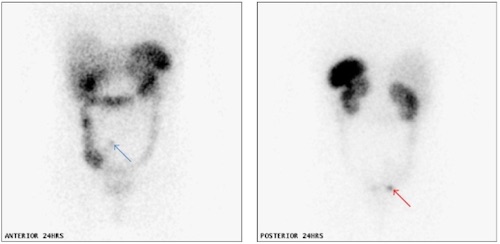
Carcinoid Tumor on Octreotide Scan 4

Carcinoid Tumor – Extent of Resection

Stomach

  • Type I/II:
    • < 1-2 cm: Endoscopic Resection
    • > 2 cm: Surgical Resection
    • Consider Antrectomy if Type I with Numerous Progressive Tumors
  • Type III: Gastrectomy (Partial vs. Total)

Small Bowel

  • Most: En Bloc Resection with Wide Lymphatic Mesenteric Clearance
    • *Some Consider Segmental Resection Alone if Single & < 1 cm with No Nodal Mets
  • Periampullary: Pancreaticoduodenectomy
  • Terminal Ileum: Right Hemicolectomy

Appendix

  • Tip & < 2 cm: Appendectomy
  • Base or ≥ 2cm: Right Hemicolectomy
    • Other Indications: Positive LN, Grade II or Mixed Histology (Goblet Cell)

Colon

  • < 1 cm: Endoscopic Polypectomy
  • > 1 cm: Partial Colectomy & Regional Lymphadenectomy

Rectum

  • High Rectum
    • < 1 cm: Endoscopic Polypectomy
    • 1-2 cm: Debated (Polypectomy vs Resection)
    • > 1 cm: Resection (LAR/APR)
  • Low Rectum
    • < 2 cm: WLE
    • > 2 cm: Resection (APR)
      • Or If Invades Muscularis Propria

References

  1. Kopáčová M, Rejchrt S, Bureš J, Tachecí I. Small intestinal tumours. Gastroenterol Res Pract. 2013;2013:702536. (License: CC BY-3.0)
  2. Pillay Y. An intestinal carcinoid causing transient jejunal intussusception in an adult-A case report. Int J Surg Case Rep. 2017;31:20-23. (License: CC BY-NC-ND-4.0)
  3. Patel CM, Sahdev A, Reznek RH. CT, MRI and PET imaging in peritoneal malignancy. Cancer Imaging. 2011 Aug 24;11(1):123-39. (License: CC BY-4.0)
  4. Sewpaul A, Bargiela D, James A, Johnson SJ, French JJ. Spontaneous Regression of a Carcinoid Tumor following Pregnancy. Case Rep Endocrinol. 2014;2014:481823. (License: CC BY-3.0)