Biliary Tract: Cholecystitis

Cholecystitis

Basics

  • Cystic Duct Obstruction
  • Initially Sterile Inflammation Until Secondarily Infected
    • Most Common Organism: E. coli

Presentation

  • RUQ Pain
    • Murphy’s Sign – Sudden “Catch” During Inspiration with Gentle RUQ Pressure
    • Boas Sign – Hyperesthesia (Increased Sensitivity) Below the Right Scapula on Back
  • Nausea & Vomiting
  • Fever
  • Leukocytosis

Diagnosis

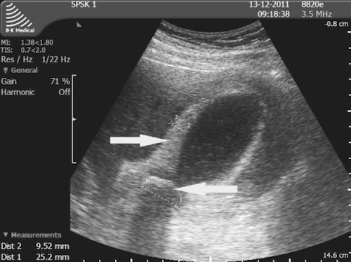
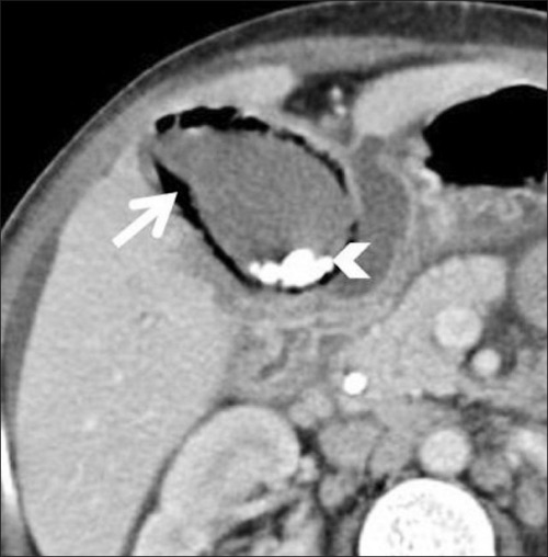
  • Diagnosis: US 95% Sensitive
  • Labs:
    • Most Sensitive Lab: CCK-Hida
    • LFT’s Normal or Only Slightly Elevated
    • Important to Rule Out Choledocholithiasis During Work-Up

Tokyo Guidelines – Severity Classification

  • Grade I (Mild): No Organ Dysfunction & Limited Disease in Gallbladder
    • Cholecystectomy Low-Risk
  • Grade II (Moderate): No Organ Dysfunction but Extensive Disease in Gallbladder
    • Cholecystectomy May be More Difficult
    • Characterized by Leukocytosis, Palpable-Tender Mass, Duration > 72 Hours & Significant Inflammation on Imaging
  • Grade III (Severe): Organ Dysfunction Present

Treatment

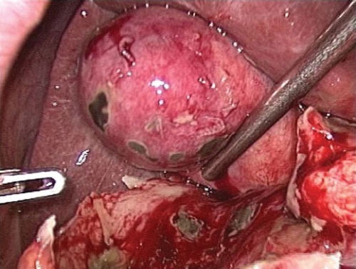
  • General Treatment: Early Cholecystectomy
  • Early (< 72 Hours) vs Late (7-45 Days) Cholecystectomy:
    • Early Has Shorter Length of Stay, Fewer Work Days Lost, Lower Total Costs & Less Wound Infection
    • Similar Complications, Conversion to Open, CBD Injury Rate and Mortality
    • No Benefit to “Cooling Off Period”
  • If Unstable or Unfit for Surgery: Percutaneous Cholecystostomy Tube
    • 90% Effective at Relieving Symptoms
    • Repeat Cholecystogram in 3-6 Weeks
      • Contrast Injected Through Catheter
      • Can Remove Catheter if Cystic Duct Patent
    • Strongly Consider Elective Interval Cholecystectomy

Pregnancy Considerations

  • First Trimester: Medical Management (NPO/ABX)
    • 85-95% Effective
    • If Fails: Percutaneous Cholecystostomy Tube as Bridge to Second Trimester Cholecystectomy
    • Surgery Risks Fetal Organogenesis
  • Second Trimester (13-26 Weeks): Cholecystectomy
  • Third Trimester: Medical Management (NPO/ABX)
    • If Fails: Percutaneous Cholecystostomy Tube as Bridge to Postpartum Cholecystectomy
    • Surgery Risks Preterm Labor

Cholecystitis with Thickened Wall on US 1

Gangrenous Gallbladder at Surgery 2

Emphysematous Cholecystitis

Basics

  • Infection by Gas-Forming Organisms
  • More Common in Diabetics & Elderly Men
  • Often Heralds Development of Gangrene, Perforation or Other Complications
  • Gallbladder Complications
    • 75% are Gangrenous
    • 20% are Perforated
  • Mortality: 25%

Organisms

  • Clostridium perfringens (GPR) – Most Common
  • E. coli – Second Most Common

Treatment

  • Stable: Emergent Cholecystectomy
  • Unstable: Cholecystostomy Tube

CT Showing Emphysematous Cholecystitis 3

Acalculous Cholecystitis

Basics

  • Gallbladder Inflammation Without Stones
  • From Biliary Stasis, Increased Viscosity & Ischemia
  • Often Present in Otherwise Ill Patients

Risk Factors

  • Trauma
  • Burns
  • Surgery
  • AIDS
  • Infection or Sepsis
  • CPR
  • TPN
  • Diabetes
  • Immunosuppression
  • Childbirth

Diagnosis

  • Diagnosis: US
  • If Uncertain: HIDA
    • Morphine Decreases False Positive Rate
    • If Critically Ill: US Sufficient, Do Not Wait for HIDA

Treatment

  • Stable: Cholecystectomy
  • If Unstable or Unfit for Surgery: Percutaneous Cholecystostomy Tube
    • If Fails: Cholecystectomy
    • Delayed Cholecystectomy Not Necessary Once Cholecystitis Resolved & Acute Illness Improves (No Stones)

References

  1. Cwik G, Skoczylas T, Wyroślak-Najs J, Wallner G. The value of percutaneous ultrasound in predicting conversion from laparoscopic to open cholecystectomy due to acute cholecystitis. Surg Endosc. 2013 Jul;27(7):2561-8. (License: CC BY-4.0)
  2. Gomes RM, Mehta NT, Varik V, Doctor NH. No 72-hour pathological boundary for safe early laparoscopic cholecystectomy in acute cholecystitis: a clinicopathological study. Ann Gastroenterol. 2013;26(4):340-345. (License: CC BY-NC-SA-3.0)
  3. Sunnapwar A, Raut AA, Nagar AM, Katre R. Emphysematous cholecystitis: Imaging findings in nine patients. Indian J Radiol Imaging. 2011 Apr;21(2):142-6. (License: CC BY-NC-SA-3.0)