Pediatric Surgery: Congenital Heart Disease

Left-to-Right Shunts

Effects

  • Oxygenated Blood Shunts from the Left-Heart to the Right-Heart
  • Causes Fluid Overload & Congestive Heart Failure
  • Signs:
    • Failure to Thrive
    • Tachypnea
    • Tachycardia
    • Hepatomegaly

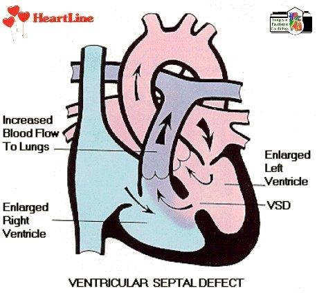
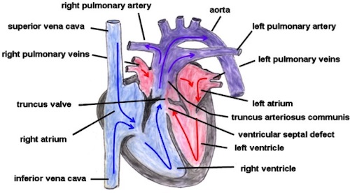
Ventricular Septal Defect (VSD)

  • Most Common Cardiac Defect
  • Blood Shunts Primarily During Systole
  • Often Born Asymptomatic but Develop Symptoms After 4-6 Weeks Due to Decreased Peripheral Vascular Resistance & Increased Shunting
  • Most Close Spontaneously in First Year
  • Treatment:
    • Small Size (< 4 mm): Medical Management
    • Medium Size (4-6 mm): Monitor & Repair at Age 5 Years (Before School Age)
    • Large Size (> 6 mm): Repair at Age 1 Year

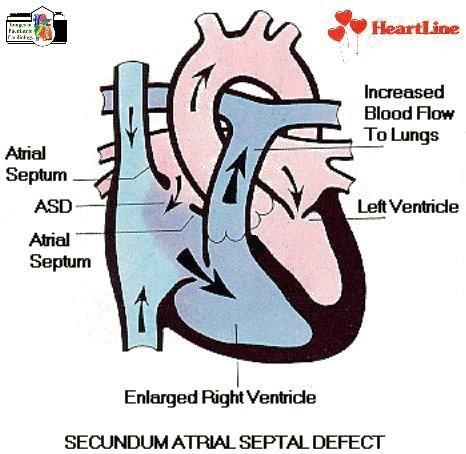
Atrial Septal Defect (ASD)

  • Third Most Common Heart Defect
  • Blood Shunts Primarily During Diastole
  • Etiology:
    • Ostium Primum Closure Defect (10%)
    • Ostium Secundum Closure Defect (80% – Most Common)
      • Also Known as Patent Foramen Ovale (PFO)
    • Sinus Venosus Defect (10%)
  • Usually Asymptomatic Until Adulthood
  • Can Have Paradoxical Emboli (DVT that Passes into Systemic Circulation through the ASD)
  • Treatment:
    • Asymptomatic: Medical Management
    • Symptomatic: Repair
      • Small-Moderate Size: Endovascular Closure
      • Large Size: Surgical Closure

Patent Ductus Arteriosus (PDA)

  • Failure of Ductus Arteriosus to Collapse After Birth
    • Allows Blood Flow from Aorta to Pulmonary Trunk
    • Normally Closes by 12-24 Hours from Increased Oxygen Tension by Ventilating Lungs
  • PDA May Be Required for Some Other Pathologies – Prostaglandin E1 Helps to Keep Patent
  • Treatment: Indomethacin or Ibuprofen (Stimulates Closure)
    • 80% Success
    • If Fails After Two Full Courses:
      • Small-Moderate Size: Endovascular Closure
      • Large Size: Surgical Closure
        • Left Posterolateral Thoracotomy
        • Close with Suture Ligation or Surgical Clips

VSD 1

ASD 1

PDA 1

Right-to-Left Shunts

Effects

  • Deoxygenated Blood Shunts from the Right-Heart to the Left-Heart
  • Causes Cyanosis
  • Spells Often Triggered by Hypovolemia or Peripheral Vasodilation (Baths)
  • Squatting May Improve Symptoms
    • Due to Increased Peripheral Vascular Resistance & Decreased Shunting

Tetralogy of Fallot (TOF)

  • Tetralogy:
    • VSD
    • Pulmonary Stenosis
    • Overriding Aorta
      • See Right Sided Deviation of Aorta
    • Right Ventricle Hypertrophy
  • Embryologic Defect: Malalignment of the Infundibular Septum
  • Treatment: Surgical Repair at 3-6 Months Old (Earlier if Symptomatic)
    • Treat Initial Spells Medically
    • β-Blockers May Reduce Spell Frequency

Eisenmenger Syndrome

  • When a Left-to-Right Shunt Becomes a Right-to-Left Shunt
  • Caused by Resulting Pulmonary Hypertension

Tetralogy of Fallot 2

Great Vessel Abnormalities

Coarctation of the Aorta

  • Narrowing of the Proximal Descending Aorta
    • Causes Increased Afterload Pressure
  • Often Associated with Other Cardiac Defects
  • Types:
    • Infantile Coarctation – Develop Symptoms within 1 Week of Life
      • Severe Obstruction Relies on PDA for Lower Body Perfusion
      • Presentation: Develops Signs of Heart Failure After Ductal Closure
    • Adult Coarctation – Develop Symptoms Later in Life
  • Diagnosis: Often Clinical in Peds
    • Echo, CT or MRI are Excellent
    • Radiology May See “Rib Notching” from Development of Large Intercostal Collaterals
  • Treatment: Surgical Correction
    • First Step: Give Prostaglandin E1 to Keep PDA Open

Coarctation of the Aorta 3

Transposition of the Great Vessels

  • Aorta & Pulmonary Artery Origins Switched
    • Results in Isolated Pulmonary & Systemic Circuits
  • Often Associated with a VSD
  • Requires PDA for Survival
  • Presentation: Cyanosis with Signs of Heart Failure
  • Treatment: Surgical Correction
    • First Step: Give Prostaglandin E1 to Keep PDA Open
    • May Also Require Initial Balloon Atrial Septostomy to Allow Additional Blood Mixing

Truncus Arteriosus

  • A Single Aorta/Pulmonary Arterial Trunk Arises from the Heart
  • Presentation: Cyanosis with Signs of Heart Failure
  • Treatment: Surgical Correction

Transposition of Great Vessels 2

Truncus Arteriosus 2

Associated Syndromes

VACTERL Syndrome

  • Associated Abnormalities:
    • Vertebral Malformations
    • Anorectal Malformations
    • Cardiac Malformations
    • Tracheoesophageal Malformations
    • Renal Malformations
    • Limb Malformations
  • Indicated Evaluation:
    • Physical Exam
    • Echocardiogram
    • Renal US
    • Sacral US

Cantrell Pentalogy

  • Cardiac Defects
  • Pericardial Defects
  • Cleft Sternum
  • Diaphragmatic Hernia
  • Omphalocele

References

  1. Grech V, Cassar J. Cardiac illustrations. Images Paediatr Cardiol. 1999 Jan;1(1):18-21.(License: CC BY-NC-SA-3.0)
  2. Salameh A, Blanke K, Daehnert I. Role of connexins in human congenital heart disease: the chicken and egg problem. Front Pharmacol. 2013 Jun 3;4:70. (License: CC BY-3.0)
  3. CDC. Wikimedia Commons. (License: Public Domain)