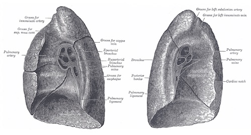
Lung Anatomy 1
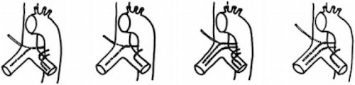
Bronchial Artery Variations; Type 1-4 (Left-to-Right) 2
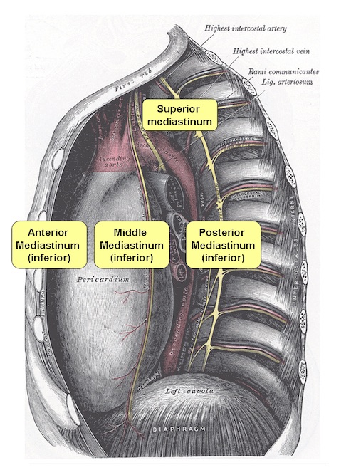
Mediastinum 1
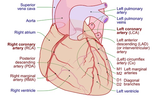
Coronary Arteries 3
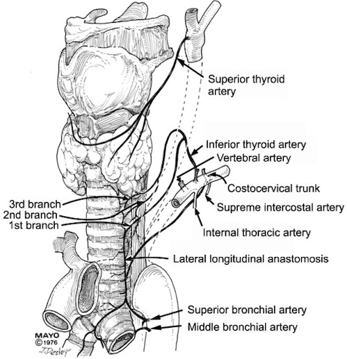
Tracheal Blood Supply 4
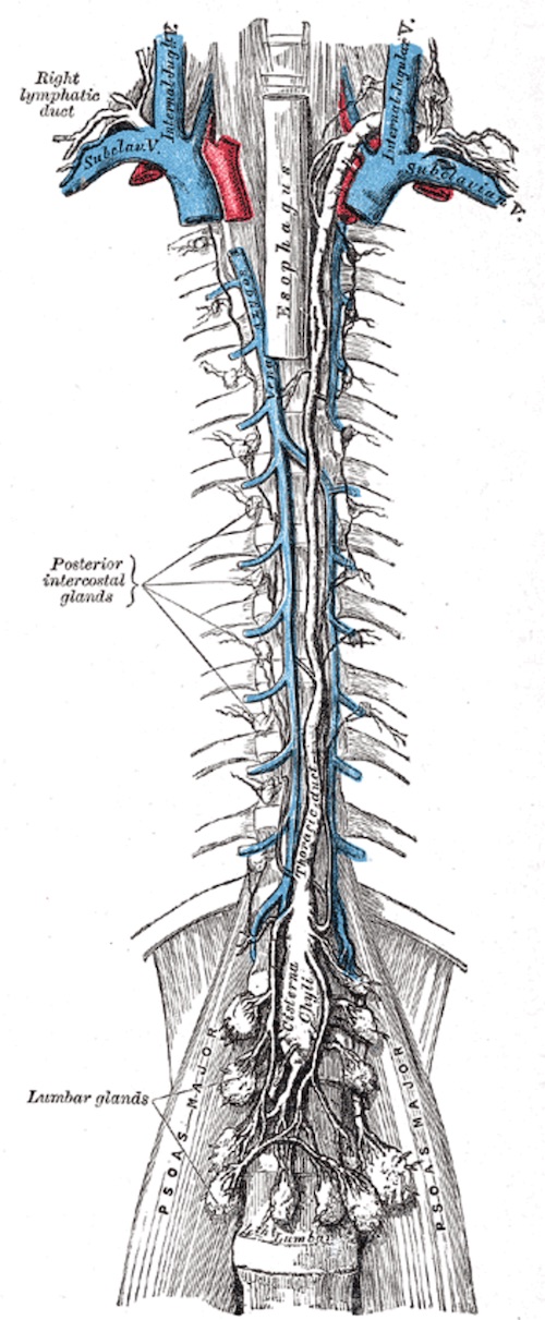
Thoracic Duct 1
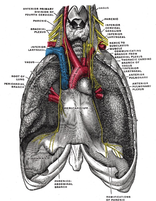
Phrenic & Vagus Nerves 1
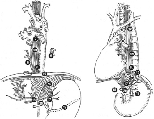
Thoracic Lymph Node Stations 5
Lung Anatomy 1
Bronchial Artery Variations; Type 1-4 (Left-to-Right) 2
Mediastinum 1
Coronary Arteries 3
Tracheal Blood Supply 4
Thoracic Duct 1
Phrenic & Vagus Nerves 1
Thoracic Lymph Node Stations 5