Esophagus: Diverticula

Zenker Diverticulum

Description

  • Location: Proximal Esophagus Through Killian’s Triangle
  • False Diverticulum
    • Does Not Involve All Layers – Only Mucosa & Submucosa
  • Pulsion Diverticulum
    • Caused by Increased Pressure from Failure of UES to Relax While Swallowing
    • Diverticulum Form at Points of Weakness
    • Most Have Associated Hiatal Hernia or GERD
  • The Most Common Esophageal Diverticulum

Symptoms

  • Dysphagia (Most Common – 80-90%)
  • Regurgitate Non-Digested Foods
  • Halitosis
  • Hoarseness
  • Cough
  • *Symptoms Worsen Throughout the Day as Diverticulum Fills with Food

Diagnosis

  • Dx: Barium Esophagram

Treatment

  • Treatment:
    • ASx & < 1 cm: Conservative Management
    • Sx or ≥ 1 cm: Repair
  • Open Surgery
    • Cricopharyngeal Myotomy & Diverticulum Repair
      • Through a Left Cervical Incision
    • Diverticulum Repair:
      • Small (< 2-3 cm): Diverticulopexy
        • Fixation to the Prevertebral Fascia
      • Moderate (3-5 cm): Diverticulopexy vs Diverticulectomy
      • Large (> 5 cm): Diverticulectomy
  • Endoscopic Diverticulotomy
    • Endoscopic Division of Septum (Including Cricopharyngeal Muscle)
    • Rigid Endoscopy Requires Neck Extension Although Newer Flexible Endoscopy Does Not
    • Previously Required At Least 2-3 cm Length Although Newer Techniques Permit Treatment of Smaller Lengths
    • Lower Complication Rates but Higher Recurrence Rate

Zenker Diverticulum

Traction Diverticulum

Description

  • Location: Mid-Esophagus
    • Usually Lateral/Peribranchial
  • True Diverticulum (Involves All Layers)
  • From Pulling at the Esophageal Wall by Scarring, Inflammation or Masses
    • Causes: Inflammation, Tuberculosis, Histoplasmosis Lymphadenopathy, Tumor

Symptoms

  • Largely ASx Until Large > 5 cm
  • Dysphagia
  • Postural Regurgitation
  • Retrosternal or Epigastric Pain
  • Heartburn
  • Cough

Diagnosis

  • Dx: Barium Esophagram
  • Consider Manometry & Upper Endoscopy
  • Possibly CT to Evaluate Paraesophageal Anatomy

Treatment

  • Treat Underlying Pathology
  • ASx: Conservative Management
  • Sx: Diverticulectomy
    • VATS Approach Preferred
    • Myotomy Not Required

Traction Diverticulum 1

Epiphrenic Diverticulum

Description

  • Location: Distal Esophagus (≤ 10 cm of GE Junction)
    • Most Common on Right Posterolateral Wall
    • Can Be Multiple & Located at Different Levels
  • Diverticulum Can Be Either True or False
  • Pulsion Diverticulum
    • Caused by Increased Pressure from a Motility Disorder
    • Diverticulum Form at Points of Weakness
    • Most Have Associated Hiatal Hernia or GERD
  • Incidence of Cancer 0.6% – Higher Risk with Delayed Diagnosis

Symptoms

  • Largely ASx Until Large > 5 cm
  • Dysphagia (Most Common)
  • Postural Regurgitation
  • Retrosternal or Epigastric Pain
  • Halitosis
  • Heartburn
  • Cough/Aspiration
  • Weight Loss

Diagnosis

  • Dx: Barium Esophagram
  • Consider Manometry & Upper Endoscopy

Treatment

  • ASx: Conservative Management
  • Sx: Diverticulectomy & Esophageal Myotomy
    • Preform Myotomy on Opposite Side to Treat Underlying Motility Disorder
    • Approaches:
      • Laparoscopy (Typically Preferred) – Consider Concurrent Fundoplication
      • Thoracotomy/VATS

Epiphrenic Diverticulum 2

References

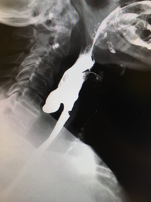
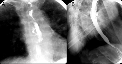
  1. Guirguis S, Azeez S, Amer S. Sarcoidosis Causing Mid-Esophageal Traction Diverticulum. ACG Case Rep J. 2016 Dec 7;3(4):e175. (License: CC BY-NC-ND-4.0)
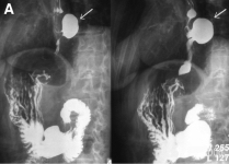
  2. Silecchia G, Casella G, Recchia CL, Bianchi E, Lomartire N. Laparoscopic transhiatal treatment of large epiphrenic esophageal diverticulum. JSLS. 2008 Jan-Mar;12(1):104-8. (License: CC BY-NC-ND-3.0)