Biliary Tract: Gallbladder Mass & Cancer

Gallbladder Nodules/Deposits

Cholesterolosis

  • Scattered Cholesterol Deposits on Wall
    • Physical Appearance of “Strawberry Gallbladder”
  • Not Premalignant
  • US: Multiple Hyperechoic & Pedunculated Masses with No Posterior Shadowing
  • Tx: Cholecystectomy

Adenomyomatosis

  • Thickened Nodule of Rokitansky-Aschoff Sinus
  • Not Premalignant (Although Recent Reports Question Pre-Malignancy)
  • Tx: Cholecystectomy

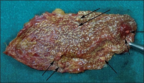
Cholesterolosis of the Gallbladder 1

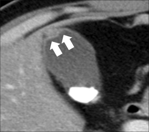
Adenomyomatosis of the Gallbladder 2

Gallbladder Polyps

Types

  • Cholesterol Polyp (Most Common)
  • Inflammatory
  • Adenoma

Malignancy Risk Factors

  • Size
    • > 1 cm (43-77% Risk)
    • > 2 cm (Nearly 100% Risk)
  • Age > 50
  • Associated Gallstones
  • Solitary Polyp (vs Multiple)

Diagnosis

  • Often Identified on US
  • Should be Evaluated with CT or MRI

Treatment

  • < 1 cm & ASx: Monitor with US
    • Cholecystectomy if: PSC/UC
  • > 1 cm or Sx: Laparoscopic Cholecystectomy
    • If > 2 cm: Consider Extended Cholecystectomy

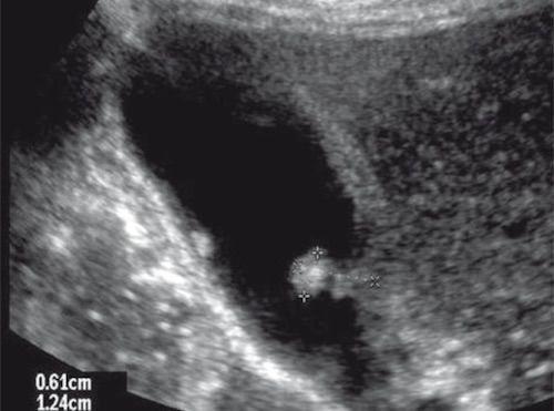
Gallbladder Polyp on US 3

Gallbladder Adenocarcinoma

Basics

  • The Most Common Biliary CA
  • Mets to Liver
  • Often Diagnosed at Advanced Stage (Poor Prognosis)
  • 5-Year Survival:
    • Overall: 5-19%
    • Early (Stage I/II): 50-64%
      • Stage I: Almost 100%
      • Stage II: 50%
    • Late (Stage III/IV): 10-24%

Risk Factors

  • Large Stones (> 3 cm) – Strongest Risk Factor
  • Female Sex
  • Polyps
  • Anomalous Pancreaticobiliary Junction
  • Chronic Infection
  • Obesity

Presentation

  • Majority (70%) Found Incidentally at Surgery for Gallstone Disease
    • Frequency: 1-2% of Cases
  • Sx: Nonspecific
  • Courvoisier Sign – Painless Palpable Gallbladder with Jaundice
    • Indicates Malignancy (Pancreas/Gallbladder)
    • *Historical Sign with Limited Utility and Many Exception

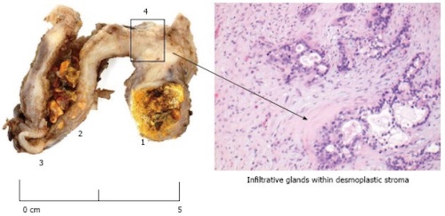
Gallbladder Carcinoma 4

TNM Staging – AJCC 8

  • TNM
  T N M
I A – Lamina Propria Invasion
B – Muscularis Invasion
1-3 LN Distant Mets
II A – Invades Perimuscular Connective Tissue on Peritoneal Side
B – Invades Perimuscular Connective Tissue on Hepatic Side
≥ 4 LN
III Invades Liver/Organs
IV Invades Portal Vein, Hepatic Artery or ≥ 2 Extrahepatic Organs
  • *Nodes Were Previously Based on Location (AJCC 7)
    • N1 Was Previously: Cystic Duct, CBD or Portal Triad LN
    • N2 Was Previously: Aortic, SMA or Celiac LN
  • Stage
  T N M
I   T1 N0 M0
II A T2a N0 M0
B T2b N0 M0
III A T3 N0 M0
B T1-3 N1 M0
IV A T4 N0-1 M0
B Any T N2 M0
Any T Any N M1

Treatment

  • If Discovered Intraoperatively: Abort Procedure & Complete Staging Prior to Return to OR
  • T1a (Confined to Mucosa): Open Cholecystectomy
    • *Some Preform Laparoscopically Although There is a Risk of Tumor Implants at Trocar Sites
    • *If Done Laparoscopic – Do Not Resect Trocar Sites (No Improved Survival)
  • ≥ T1b (Invades Muscle): Extended Cholecystectomy & Portal Lymphadenectomy
    • Resection:
      • Formal Segment IVb & V Anatomic Liver Resection – Historical Preference Now Fallen Out of Favor
      • Extended Cholecystectomy – Nonanatomic Resection Extending into the Gallbladder Fossa
    • Portal Lymphadenectomy Should Harvest ≥ 6 Lymph Nodes
    • If Found on Postoperative Pathology: Return to OR & Complete Resection
    • If Extends to CBD or Positive Cystic Duct Margin (Frozen Section): CBD Resection & Hepaticojejunostomy
  • Unresectable or Mets: Chemotherapy
    • Surgery Absolute Contraindications:
      • Aortic, SMA or Celiac LN
      • Distant Metastases
      • Malignant Ascites
      • Encasement of Major Vessels

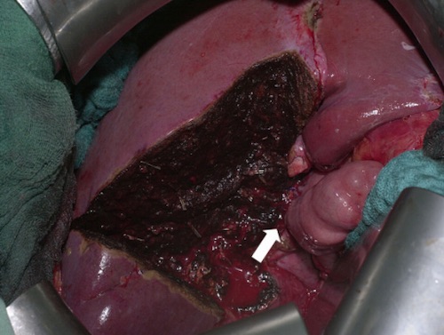
Formal Segment IVb & V Liver Resection 5

References

  1. Yadav S, Jategaonkar P, Bijlani M. Gallbladder polyps: an ambiguous cause of biliary colic. Ann Med Health Sci Res. 2014 Sep;4(Suppl 3):S332-3. (License: CC BY-NC-SA-3.0)
  2. Kim BS, Oh JY, Nam KJ, Cho JH, Kwon HJ, Yoon SK, Jeong JS, Noh MH. Focal thickening at the fundus of the gallbladder: computed tomography differentiation of fundal type adenomyomatosis and localized chronic cholecystitis. Gut Liver. 2014 Mar;8(2):219-23. (License: CC BY-NC-3.0)
  3. Walas MK, Skoczylas K, Gierbliński I. Standards of the Polish Ultrasound Society – update. The liver, gallbladder and bile ducts examinations. J Ultrason. 2012 Dec;12(51):428-45. (License: CC BY-NC-ND-3.0)
  4. Patel K, Dajani K, Iype S, Chatzizacharias NA, Vickramarajah S, Singh P, Davies S, Brais R, Liau SS, Harper S, Jah A, Praseedom RK, Huguet EL. Incidental non-benign gallbladder histopathology after cholecystectomy in an United Kingdom population: Need for routine histological analysis? World J Gastrointest Surg. 2016 Oct 27;8(10):685-692. (License: CC BY-NC-4.0)
  5. Scheingraber S, Justinger C, Stremovskaia T, Weinrich M, Igna D, Schilling MK. The standardized surgical approach improves outcome of gallbladder cancer. World J Surg Oncol. 2007 May 21;5:55. (License: CC BY-2.0)