Small Intestine: Internal Hernia

Definitions

Definition

  • Herniation Through an Opening in the Peritoneum or Mesentery
  • Opening Can Be Congenital or Acquired

Types

  • Paraduodenal
    • Historically the Most Common (53%) – Not Anymore Given Frequency of Newer Laparoscopic Procedures
  • Foramen of Winslow
  • Peri-Cecal: Herniation in the Pericecal Fossa
  • Inter-Sigmoid: Herniation Through the Intersigmoid Fossa (Often Easily Reducible)
    • Debated if it is a True Aperture/Hernia
  • Supravesical: Herniation into the Space of Retzius (Can Be Anterior or Posterior to the Bladder)
  • Trans-Omental: Herniation Through the Greater or Lesser Omentum
  • Trans-Mesenteric: Herniation Through a Complete Defect in the Mesentery
    • Either from a Congenital Defect or from a Surgical Defect (Retrocolic Roux-en-Y)
  • Retro-Anastomotic: Related to a Surgical Anastomosis
    • Risk After Roux-en-Y Gastric Bypass: 3-5%
    • Risk After Laparoscopic Colorectal Surgery: 0.65%

Paraduodenal Hernias

  • Left Paraduodenal Hernia: Herniation Through Landzert’s Fossa
    • Most Common (75%)
  • Right Paraduodenal Hernia: Herniation Through Waldeyer’s Fossa

Sigmoid-Related Hernias

  • Inter-Sigmoid: Herniation Through the Inter-Sigmoid Fossa (Often Easily Reducible)
    • Debated if it is a True Aperture/Hernia
  • Trans-Mesosigmoid: Herniation Through a Complete Defect in the Sigmoid Mesocolon
  • Intra-Mesosigmoid: Herniation into an Incomplete Defect in the Sigmoid Mesocolon Through Only a Single Layer with the Sac in the Sigmoid Mesocolon (Most Commonly the Left)

Mesenteric Defects After a Roux-en-Y Gastric Bypass

  • Petersen’s Space – Mesenteric Defect Between the Roux Limb Mesentery & Transverse Mesocolon
  • Brolin’s Space (Mesojejunal Window) – Mesenteric Defect at the Jejunojejunostomy
  • Transverse Mesocolon Defect
    • Only Present After Retrocolic Procedures

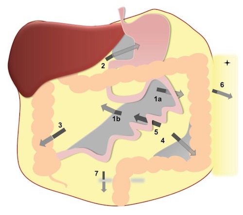
Sites of Internal Hernia: Left Paraduodenal Hernia (1a); Right Paraduodenal Hernia (1b); Foramen of Winslow Hernia (2); Pericecal Hernia (3); Sigmoid-Related Hernia (4); Transmesenteric Hernia (5); Transomental Hernia (6); Supravesical Hernia (7) 1

Left Paraduodenal Hernia 2

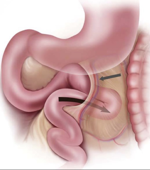
Right Paraduodenal Hernia; (A) Normal Anatomy, (B) Right Paraduodenal Hernia 3

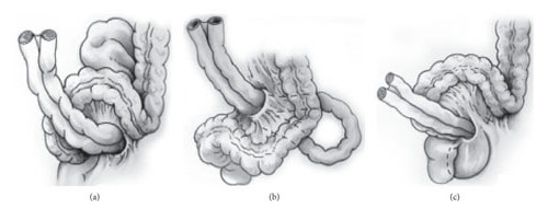
Sigmoid-Related Hernias; (a) Intersigmoid, (b) Trans-Mesosigmoid, (c) Intra-Mesosigmoid 4

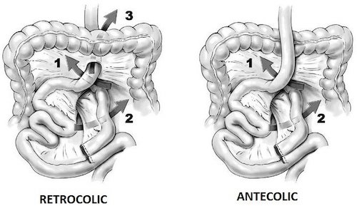
Mesenteric Defects After Roux-en-Y Gastric Bypass; (1) Petersen’s Space, (2) Brolin’s Space, (3) Transverse Mesocolic Window (Only in Retrocolic Bypass) 5

Congenital Anatomic Defects

Landzert’s Fossa

  • Present in 2% of the Population
  • Located Behind the Fourth Portion of the Duodenum
  • Formed by the Mesenteric Fold from the IMV & Left Colic Artery

Waldeyer’s Fossa

  • Present in < 1% of the Population
  • Located Under the Third Portion of the Duodenum
  • Formed by the Mesenteric Fold from the SMA

Foramen of Winslow (Epiploic/Omental Foramen)

  • Communication Between the Greater Sac & Lesser Sac
  • Anteriorly Bordered by the Hepatoduodenal Ligament

Pericecal Fossa

  • Located Behind the Cecum/Ascending Colon
  • 4-Subtypes: Ileocolic, Ileocecal, Retrocecal, & Paracecal

Intersigmoid Fossa

  • Present in 65% of the Population
  • Formed Between 2 Adjacent Sigmoid Segments & Their Mesentery
  • Debated if it is a True Aperture

Supravesical Fossa

  • Triangular Area Bounded by the Peritoneal Reflection Over the Dome of Bladder & Lateral Umbilical Folds
  • Space of Retzius: Potential Space Below the Supravesical Fossa; Between Pubic Symphysis and Bladder

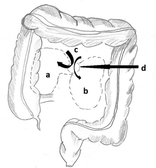
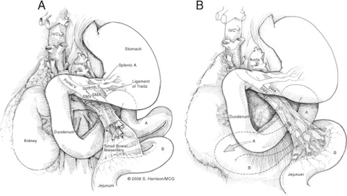
Paraduodenal Hernia Fossae; (a) Waldeyer’s Fossa, (b) Landzert’s Fossa, (c) Retroperitoneum, (d) Hernial Orifice 6

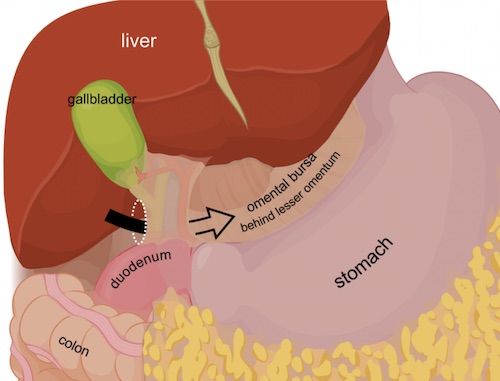
Foramen of Winslow 7
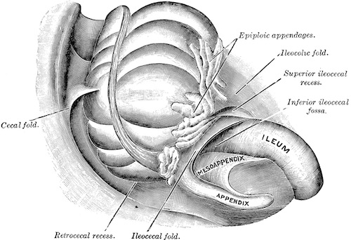
Pericecal Fossa 8

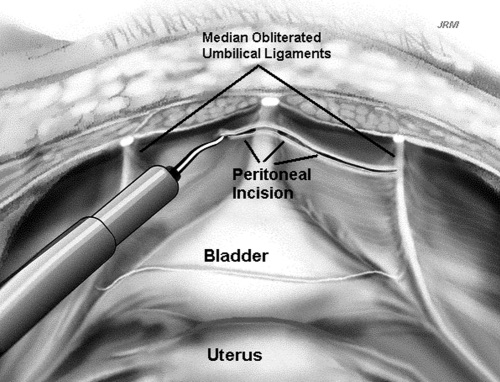
Incision into the Space of Retzius 9

Management

Presentation

  • Can Be Asymptomatic
  • Small Bowel Obstruction (SBO)
  • Chronic Postprandial Pain
  • Bowel Ischemia/Strangulation

Diagnosis

  • Primarily Made by CT
  • CT Findings:
    • Small Bowel Obstruction
    • Closed-Loop Obstruction
    • Mesenteric “Swirling”
    • Small Bowel Herniation into an Abnormal Location – Requires Sufficient Knowledge of Anatomic Spaces
  • May Be Found Intraoperatively

Treatment

  • Surgical Repair

Peterson’s Hernia; (A) AXR Showing SBO, (B) CT Showing Mesenteric Swirl Sign 10

References

  1. Lanzetta MM, Masserelli A, Addeo G, Cozzi D, Maggialetti N, Danti G, Bartolini L, Pradella S, Giovagnoni A, Miele V. Internal hernias: a difficult diagnostic challenge. Review of CT signs and clinical findings. Acta Biomed. 2019 Apr 24;90(5-S):20-37. (License: CC BY-4.0)
  2. Hassani KI, Aggouri Y, Laalim SA, Toughrai I, Mazaz K. Left paraduodenal hernia: A rare cause of acute abdomen. Pan Afr Med J. 2014 Mar 27;17:230. (License: CC BY-2.0)
  3. Bittner JG 4th, Edwards MA, Harrison SJ, Li K, Karmin PN, Mellinger JD. Laparoscopic repair of a right paraduodenal hernia. JSLS. 2009 Apr-Jun;13(2):242-9. (License: CC BY-NC-ND-3.0)
  4. Chiarini S, Ruscelli P, Cirocchi R, D’Andrea V, Sensi B, Santoro A, Corsi A, Zepponi F, Fedeli P, Gioia S. Intersigmoid Hernia: A Forgotten Diagnosis-A Systematic Review of the Literature over Anatomical, Diagnostic, Surgical, and Medicolegal Aspects. Emerg Med Int. 2020 Jun 1;2020:4891796. (License: CC BY-4.0)
  5. Kim, Y., & Crookes, P. F. (2014). Complications of Bariatric Surgery. In (Ed.), Essentials and Controversies in Bariatric Surgery. IntechOpen. (License: CC BY-3.0)
  6. Mehra R, Pujahari AK. Right paraduodenal hernia: report of two cases and review of literature. Gastroenterol Rep (Oxf). 2016 May;4(2):168-71. (License: CC BY-4.0)
  7. Wikimedia Commons. (License: CC BY-SA-3.0)
  8. Gray H. Anatomy of the Human Body (1918). Public Domain.
  9. Pikaart DP, Miklos JR, Moore RD. Laparoscopic removal of pubovaginal polypropylene tension-free tape slings. JSLS. 2006 Apr-Jun;10(2):220-5. (License: CC BY-NC-ND-3.0)
  10. Jang JS, Shin DG. A Peterson’s hernia and subsequent small bowel volvulus: surgical reconstruction utilizing transverse colon as a new Roux-en-Y limb – 1 case. J Korean Surg Soc. 2013 Dec;85(6):309-13. (License: CC BY-NC-3.0)