Pediatric Surgery: Intestinal Atresia

Duodenal Atresia

Basics

  • Blockage Due to Duodenal Malformation
  • Cause Complete (81%) or Partial (19%) Obstruction
  • Most Common (85%) at the Junction of First/Second Portions
  • Most Common Cause of Duodenal Obstruction in Newborns < 1 Week

Cause

  • Cause: Failure of Recanalization
  • Normal Development
    • Week 6-7: Occlusion as Epithelium Proliferates
    • Week 8-10: Patency Restored

Associations

  • Half (55%) Have Other Congenital Anomalies
  • Down Syndrome (28.2% – Most Common)
  • Annular Pancreas (23.1%)
  • Congenital Heart Disease (22.6%)
  • Malrotation (19.7%)
  • Esophageal Atresia (8.5%)

Types

  • Type 1 (92%) – Obstructing Septum/Web of Mucosa & Submucosa
    • Intact Muscularis, Bowel Wall & Mesentery
  • Type 2 (1%) – A Short Fibrous Cord Connects Two Blind Ends
    • Intact Mesentery
  • Type 3 (7%) – No Connection Between Two Blind Ends with a V-Shaped Mesenteric Defect

Presentation

  • Bilious Emesis
  • Abdomen Nondistended – May See Upper Abdominal Fullness
  • Results in Microcolon
  • Maternal Polyhydramnios

Diagnosis

  • Prenatal US
  • Abdominal XR – Classic “Double Bubble
    • May Require Injection of 30-60 cc Air to Demonstrate Double Bubble
    • No Distal Gas
  • Upper GI Study – Required to Rule Out Malrotation/Volvulus

Treatment

  • Initial Management: NG/OG Tube Decompression, IV Fluids & TPN
    • Echocardiogram First to Rule out Cardiac Defects
  • Definitive Management: “Diamond” Duodenoduodenostomy
    • If Obstruction Due to Only A Thin Web: Consider Web Excision Rather than Bypass
    • If Proximal Bowel is Severely Dilated: Consider Tapering Duodenoplasty
      • Faster Return of Normal Peristalsis
    • If Duodenum-Duodenum Anastomosis Anatomically Unfeasible: Duodenojejunostomy (#2) or Gastrojejunostomy (#3)

Diamond Duodenoduodenostomy

  • Incisions:
    • Proximal Transverse Incision (More Dilated)
    • Distal Longitudinal Incision
  • Midpoints of Each Incision Joined to the Endpoints of the Other
  • Keeps Itself Patent to Allow Earlier Transit

Duodenal Atresia 1

Duodenal Atresia, “Double Bubble” on XR 2

Diamond Duodenoduodenostomy 3

Jejunoileal Atresia

Basics

  • Most Common Site of Atresia
  • Most Common Congenital Anomaly of the Small Intestine
  • 25-35% Have Other Congenital Anomalies
    • Less Common Than Duodenal Atresia
  • Most Are a Single Event Although 6-20% Have Multiple Atresias
  • Risk for Dysfunctional Proximal Peristalsis
    • Due to Intestinal Smooth Muscle Hypertrophy, Hypoplasia of Intramural Nerves & Decreased Number of Interstitial Cells of Cajal
  • Most Common Cause of Early Death: Infection (PNA/Peritonitis/Sepsis)

Cause

  • Cause: Intrauterine Vascular Accidents
  • Risk Factors:
    • Cocaine
    • Pseudoephedrine
    • Ergotamine Tartrate or Caffeine in Management of Migraines

Presentation

  • Bilious Emesis
  • Abdomen Distended
  • Failure to Pass Meconium
  • Results in Microcolon
  • Maternal Polyhydramnios
  • Jaundice

Jejunal Atresia 4

Types

  • Louw & Barnard Classification:
    • Type I (19%) – Mucosal Atresia (Intact Muscularis, Bowel Wall & Mesentery)
    • Type II (31%) – Blind Ends (Intact Mesentery)
    • Type III (46%) – Blind Ends with V-Shaped Mesenteric Defect
  • Contemporary Classification:
    • Type I: Mucosal Atresia (Intact Muscularis, Bowel Wall & Mesentery)
    • Type II: Blind Ends (Intact Mesentery)
    • Type IIIa: Blind Ends with V-Shaped Mesenteric Defect
    • Type IIIb: Apple-Peel/Christmas-Tree Atresia
    • Type IV: Multiple Atresias

Diagnosis

  • Prenatal US
    • Better at Detecting Proximal Atresia Than Distal
  • Abdominal XR
  • Upper GI Study

Treatment

  • Initial Management: NG/OG Tube Decompression, IV Fluids & TPN
  • Definitive Management: End-to-End Anastomosis
    • Resect the Distal End of the Proximal Segment (Smooth Muscle Hypertrophy & Decreased Interstitial Cells of Cajal Cause a Functional Obstruction)
    • Side-to-Side Risks Functional Obstruction & Blind Loop Syndrome
  • Options if Large Size Difference Occurs:
    • Larger End: Tapering Enteroplasty
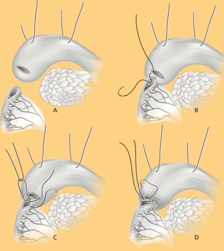
    • Smaller End: Cheatle Slit (Longitudinal Incision on Antimesenteric Border for Wider Anastomosis)
  • If Concerned for Short Bowel Syndrome: Consider G-Tube Placement

References

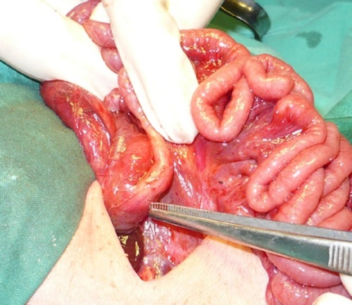
  1. Mirza B, Sheikh A. Multiple associated anomalies in patients of duodenal atresia: a case series. J Neonatal Surg. 2012 Apr 1;1(2):23. (License: CC BY-3.0)
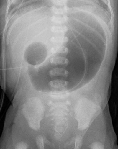
  2. Klinikum Stuttgart KO. Wikimedia Commons. (License: CC BY-SA-3.0)
  3. van der Zee DC. Laparoscopic repair of duodenal atresia: revisited. World J Surg. 2011 Aug;35(8):1781-4. (License: CC BY-NC-2.0)
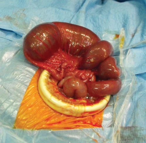
  4. Federici S, Sabatino MD, Domenichelli V, Straziuso S. Worst Prognosis in the “Complex” Jejunoileal Atresia: Is It Real? European J Pediatr Surg Rep. 2015 Jun;3(1):7-11. (License: CC BY-NC-SA-4.0)