Liver: Mass CT Characteristics

CT Characteristics

Simple Cyst

  • Well-Demarcated
  • No Enhancement
  • No Visible Wall

Mucinous Cystic Neoplasm (MCN)

  • Unilocular or Multilocular with Septations
  • Thickened Irregular Wall

Hemangioma

  • Arterial Phase: Peripheral Enhancement
  • Portal Venous Phase: Centripetal Flow
  • Delayed Phase: Isodense Then Hypodense
  • MRI:
    • Smooth, Well-Demarcated & Homogenous
    • Low Signal Intensity on T1
    • Peripheral Nodular Enhancement

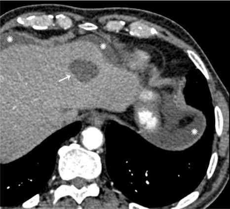
Simple Cyst of the Liver 1

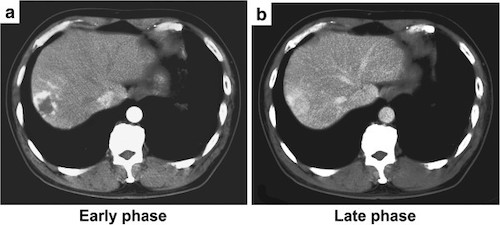
Hepatic Hemangioma. (a) Early Arterial Peripheral Enhancement, (b) Late Homogenous Attenuation 2

Pyogenic Abscess

  • Well-Defined
  • Central Hypoattenuation
  • Rim Enhancement with Peripheral Edema

Amebic Abscess

  • In Peripheral Liver
  • No Rim Enhancement
  • Peripheral Edema

Hydatid Cyst

  • Inner Wall Infoldings
  • Double-Walled with Calcified Rim
  • May See Septa/Daughter Cysts

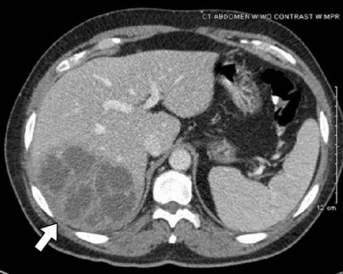
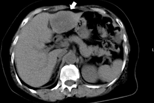
Pyogenic Liver Abscess 3
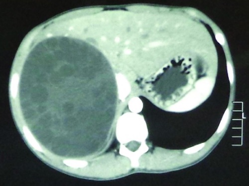
Amebic Abscess 4

Hydatid Cyst of the Liver 5

Focal Nodular Hyperplasia (FNH)

  • Arterial Phase: Homogenous Enhancement
  • Portal Venous Phase: Precontrast Density
  • Central Stellate Scar
  • Liver Scan: Hot

Hepatocellular Adenoma

  • Well Circumscribed
  • Variable Attenuation Depending on Calcification, Hemorrhage (Hyperattenuating) or Fat Content (Hypoattenuating)
  • Similar to FNH
  • Arterial Phase: Homogenous Enhancement
  • Portal Venous Phase: Precontrast Density
  • Liver Scan: Cold

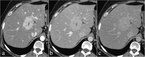
FNH. (a) Early Arterial Homogenous Enhancement with Central Scar, (b) Portal Venous Washout, (c) Delayed Phase Isodensity 6

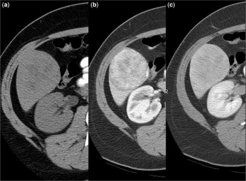
Hepatic Adenoma. (a) Precontrast, (b) Enhancement on Arterial Phase, (c) Gradual Washout on Delayed Phase 7

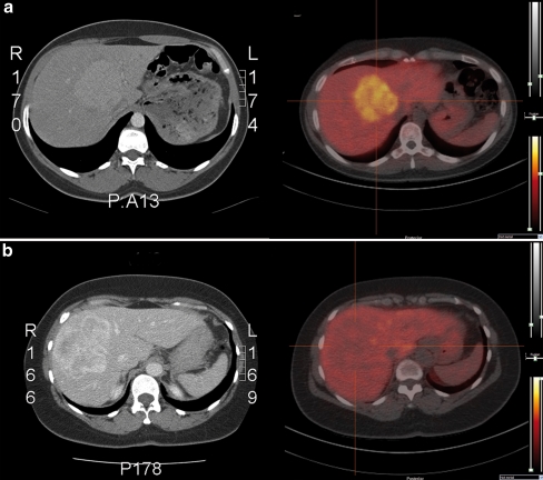
Liver Scan. (a) FNH (Hot), (b) HCA (Cold) 8

Hepatocellular Carcinoma (HCC)

  • Arterial Phase: Early Enhancement
  • Portal Venous/Delayed Phase: Washout
  • Liver Scan: Cold

HCC FLC

  • Arterial Phase: No Enhancement
  • May Have Central Scar

Metastases

  • Arterial Phase: No Enhancement
    • Some Such as NET, RCC, etc. May Have Rapid Enhancement

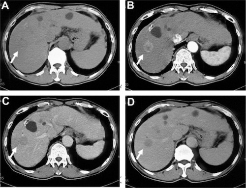
Hepatocellular Carcinoma. (a) Noncontrast, (b) Arterial Phase Enhancement, (c) Portal Phase Washout, (d) Delayed Phase Hypoattenuation 9

Liver Metastasis from Colon Cancer 10

CT Contrast Phases

Early Systemic Arterial Phase

  • Time from Injection: 15-20 Seconds
  • Target: Arteries

Late Systemic Arterial Phase (Arterial Phase)

  • Also Referred to as Early Portal Venous Phase
  • Time from Injection: 35-40 Seconds
  • Target: Structures with Arterial Blood Supply

Hepatic Phase

  • Also Referred to as Late Portal Venous Phase
  • Time from Injection: 70-80 Seconds
  • Target: Liver Parenchyma & Structures with Portal Vein Supply

Systemic Venous Phase

  • Time from Injection: 180 Seconds
  • Target: Venous Thrombosis

Delayed/Washout Phase

  • Time from Injection: 6-15 Minutes
  • Target: Disappearance of All Contrast Unless Tissue Fibrotic

CT Characteristic Comparison

Non-Solid Liver Mass CT Characteristics

  Wall/Rim Periphery Extra
Simple Cyst None
MCN Thickened & Irregular Septations
Hydatid Cyst Double-Walled & Calcified Rim
Pyogenic Abscess Rim Enhancement Edema
Amebic Abscess No Rim Enhancement Edema

Solid Liver Mass CT Characteristics

  Arterial Phase Portal Venous Phase Extra
Hemangioma Peripheral Centripetal Hypodense Delayed Phase
FNH Homogenous Precontrast Central Stellate Scar Hot Liver Scan
Adenoma Homogenous Precontrast Variable Attenuation
HCC Early Enhancement Washout
HCC FLC No Enhancement May Have Scar
Metastases No Enhancement

References

  1. Lazoura O, Vassiou K, Kanavou T, Vlychou M, Arvanitis DL, Fezoulidis IV. Incidental non-cardiac findings of a coronary angiography with a 128-slice multi-detector CT scanner: should we only concentrate on the heart? Korean J Radiol. 2010 Jan-Feb;11(1):60-8. (License: CC BY-NC-3.0)
  2. Shimada Y, Takahashi Y, Iguchi H, Yamazaki H, Tsunoda H, Watanabe M, Oda M, Yokomori H. A hepatic sclerosed hemangioma with significant morphological change over a period of 10 years: a case report. J Med Case Rep. 2013 May 28;7:139. (License: CC BY-2.0)
  3. Livingston LV, Perez-Colon E. Streptococcus intermedius Bacteremia and Liver Abscess following a Routine Dental Cleaning. Case Rep Infect Dis. 2014;2014:954046. (License: CC BY-3.0)
  4. Garvin KW, Willig JH. Amebic liver abscess. Am J Trop Med Hyg. 2010 Nov;83(5):961. (License: CC BY-2.5)
  5. Ma Z, Yang W, Yao Y, Liu Q. The adventitia resection in treatment of liver hydatid cyst: a case report of a 15-year-old boy. Case Rep Surg. 2014;2014:123149. (License: CC BY-3.0)
  6. Scialpi M, Pierotti L, Gravante S, Piscioli I, Pusiol T, Schiavone R, D’Andrea A. Split-bolus versus triphasic multidetector-row computed tomography technique in the diagnosis of hepatic focal nodular hyperplasia: a case report. J Med Case Rep. 2014 Dec 14;8:425. (License: CC BY-4.0)
  7. Gore RM, Thakrar KH, Wenzke DR, Newmark GM, Mehta UK, Berlin JW. That liver lesion on MDCT in the oncology patient: is it important? Cancer Imaging. 2012 Sep 28;12(2):373-84.(License: CC BY-4.0)
  8. van den Esschert JW, Bieze M, Beuers UH, van Gulik TM, Bennink RJ. Differentiation of hepatocellular adenoma and focal nodular hyperplasia using 18F-fluorocholine PET/CT. Eur J Nucl Med Mol Imaging. 2011 Mar;38(3):436-40. (License: CC BY-NC-2.0)
  9. Murakami T, Tsurusaki M, Hyodo T, Imai Y. Clinical utility of imaging for evaluation of hepatocellular carcinoma. J Hepatocell Carcinoma. 2014 Jul 14;1:101-8. (License: CC BY-NC-3.0)
  10. Ito T, Kushida T, Sakurada M, Maekawa H, Orita H, Mizuguchi K, Sato K. Two cases of laparoscopic simultaneous resection of colorectal cancer and synchronous liver metastases in elderly patients. Int J Surg Case Rep. 2016;26:134-7. (License: CC BY-NC-ND-4.0)