Urolithiasis: Stone in Any Part of the Urinary Tract
Nephrolithiasis: Kidney Stones or Renal Calculi
*Often Used to Broadly Refer to Any Stone in the Urinary Tract
Ureterolithiasis: Stone in the Ureter
Stone Types
Radiopaque Stones:
Calcium Oxalate (70-80% – Most Common Stone)
Calcium Phosphate (15%)
Struvite (Mg Ammonium Phosphate) (1%)
Staghorn Calculi Within Pelvis
From Proteus mirabilis Infection (Produce Urease)
Radiolucent Stones:
Uric Acid (8%)
Cysteine (1-2%)
From Congenital Disorders of Cysteine Reabsorption
Risk Factors
Older Age
Male Sex
White Ethnicity
Functional Disability
Prior Stones
Recurrence Risk: 10-30% at 3-5 Years, 50% at 10 Years
Specific Risk Factors for Calcium Oxalate Stones:
Low Urine Output
High Urine Calcium or Oxalate
High Urine pH
Low Urine Citrate
Primary Hyperparathyroidism
Diabetes
Increased GI Absorption of Oxalate in Colon – Due to IBD, Short Gut Syndrome or Bowel Resection)
Betel Quid Chewing (Practice in Southeast Asia)
Specific Risk Factors for Uric Acid Stones:
Gout
Obesity
Low Urine pH
Myeloproliferative Disorders
Presentation
Colicky Pain – Severe Episodes Lasting 20-60 Minutes
Hematuria
Nausea & Vomiting
Dysuria
Urinary Urgency
Complications
Urinary Obstruction & Hydronephrosis
Renal Injury/Failure
Urinary Tract Infection/Pyelonephritis
Diagnosis
Primary Diagnosis: CT (Without Contrast)
Other Options: MRI or IV Pyelography
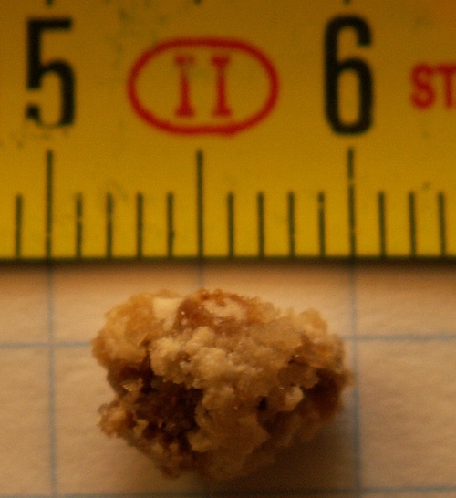
Nephrolithiasis 1
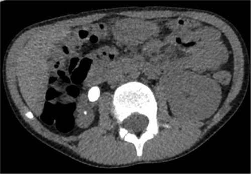
Large Right Ureterolithiasis on CT 2
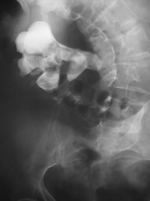
Staghorn Nephrolithiasis on Abdominal XR 3
Treatment
Medical Treatment
Primary Tx: Pain Control (NSAIDs Preferred)
Tamsulosin (Alpha Blocker) for Stones 5-10 mm in Diameter
Emergent Surgery
Indications:
Obstructing Stone & UTI/Sepsis
Bilateral Obstruction & AKI
Obstruction & AKI with a Solitary Functional Kidney
Urgent Decompression is Preferred Approach
Generally Wait to Remove Stone Until Infection is Cleared
Stone Manipulation in Setting of UTI Can Cause Severe Sepsis
Surgical Approach:
Ureteroscopy with Stent Placement
Percutaneous Nephrolithotomy with Percutaneous Nephrostomy Tube
Elective Surgery
Indications:
Severe Symptoms
Diameter > 10 mm
Medical Failure After 4-6 Weeks
Persistent Obstruction Due to Stones
Recurrent UTI Due to Stones
Surgical Options:
Ureteroscopy – Often the Preferred Approach
May Leave a Ureteral Stent After Stone Excision
Extra-Corporeal Shock Wave Lithotripsy (ESWL) – May Be Considered for Small-Medium Sized Stones in the Proximal-Mid Ureter
Percutaneous Nephrolithotomy – Generally Reserved as Second-Line for Large/Complex Stones
May Leave a Percutaneous Nephrostomy Tube After Stone Excision
Surgical Excision (Laparoscopic, Robotic or Open) – Rarely Used
References
Wal RR. Wikimedia Commons. (License: Public Domain)
Patel C, Modgil V, Luscombe C, Liu S. A unique presentation, and management, of acute urinary retention in a young boy with underlying vesicoureteral reflux. J Surg Case Rep. 2013 Sep 7;2013(9):rjt047. (License: CC BY-NC-3.0)
Dilmen N. Wikimedia Commons. (License: CC BY-SA-3.0)