Vascular: Other Venous Emboli

Fat Emboli

Definitions

  • Fat Embolism – Fat Globules in the Pulmonary Circulation
  • Fat Embolism Syndrome (FES) – Clinical Syndrome that Develops After a Fat Embolism
    • From Fat Globule Release into Circulation
    • Most Fully Resolve Spontaneously within a Few Days

Causes

  • Orthopedic Fractures
    • Long Bone (Femur) & Pelvic Fractures – Accounts for Almost All Cases
    • High Content of Fat in Bone Marrow
    • Typically Present 1-3 Days After Fractures
  • Trauma
  • Burns
  • Pancreatitis
  • CPR

Presentation

  • Classic Symptomatic Triad:
    • Hypoxemia – Generally the First Sign
    • Neurologic Abnormalities – Generally Develops After Hypoxemia
    • Petechial Rash – Generally the Last to Develop
  • Pulmonary Signs:
    • Hypoxemia – The Most Common Feature (> 90%)
    • Dyspnea
    • Tachypnea
    • Often Develop a Syndrome Indistinguishable from ARDS
  • Neurologic Signs:
    • Altered Mental Status
    • Seizures
    • Focal Deficits
  • Petechial Rash (20-50%) – From Fat Occlusion of Dermal Capillaries
  • Less Common Manifestations:
    • Anemia
    • Fever
    • DIC
    • Hypotension
    • Myocardial Depression

Diagnosis

  • Primary Diagnosis: Clinical Diagnosis
  • CTA Can Rule Out PE
  • Supportive Findings (Low Sensitivity/Specificity – Rarely Performed):
    • Bronchoscopy with Bronchioalveolar Lavage (BAL) – Detects Fat Droplets in Alveolar Macrophages
    • Pulmonary Artery Catheter – Fat Detected in a Wedge Sample
    • Urine/Sputum Samples – Detection of Fat

Treatment

  • Primary Treatment: Supportive
  • Early Fracture Immobilization/Repair Prevents FES – Unclear if it Helps Treat Established FES
  • Therapeutic Anticoagulation Not Advised – Risk for Hemorrhage & Increased Free Fatty Acid Production

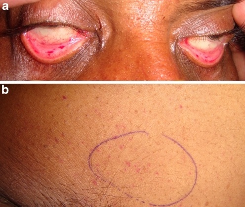
FES Petechial Rash 1

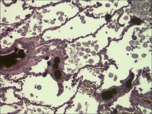
Lung Section with Extensive Fat Embolism and Fat Droplets in Alveolar Macrophages 2

Air Embolism

Causes

  • Central Venous Catheter – Most Common Cause
  • Surgery
  • Vascular Interventions
  • Trauma
  • Mechanical Ventilation Barotrauma

Presentation

  • Hypoxemia & Respiratory Distress
  • Neurologic Abnormalities if Embolized to Arterial System
  • Millwheel” Murmur: Loud Churning
  • Myocardial Infarction is Rare – Usually Due to Air Entrance into the Arterial System Through a Congenital Heart Defect

Diagnosis

  • Diagnosis: Demonstration of Intravascular Air with Known Risk Factor
    • Often Rapidly Absorbed Prior to Imaging
    • Consider Echo or CT

Treatment

  • Primary Treatment: Repositioning & Supportive Cares (Supplemental Oxygen)
  • Repositioning:
    • “Durant’s Maneuver”Left Lateral Decubitus (Left Down)
    • TrendelenburgHead Down
    • Goal: Positioning to Trap Air in the Right Ventricle
    • First Step is Repositioning Even Before Diagnosis if There is Concern
  • Options If Hemodynamically Unstable:
    • Hyperbaric Oxygen
    • Attempt Aspiration Through a Central Venous Catheter
    • Chest Compressions to Force Air into Smaller Vessels

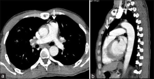
Air Embolism (Arrow) in Pulmonary Artery 3
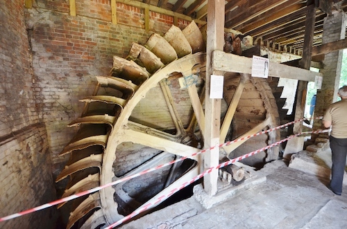
Mill Wheel 1

References

  1. Sogunuru G, Moka N. Fat embolism syndrome. Int J Emerg Med. 2010 Apr 1;3(4):471. (License: CC BY-NC-2.0)
  2. Eriksson EA, Schultz SE, Cohle SD, Post KW. Cerebral fat embolism without intracardiac shunt: A novel presentation. J Emerg Trauma Shock. 2011 Apr;4(2):309-12. (License: CC BY-NC-SA-3.0)
  3. Sodhi KS, Saxena AK, Chandrashekhar G, Bhatia A, Singhi S, Agarwal R, Khandelwal N. Vascular air embolism after contrast administration on 64 row multiple detector computed tomography: A prospective analysis. Lung India. 2015 May-Jun;32(3):216-9. (License: CC BY-NC-SA-3.0)
  4. Dace A. Wikimedia Commons. (License: CC BY-SA-2.0)