Parapneumonic Effusion – Pleural Fluid that Develops in Response to Adjacent Pneumonia
Uncomplicated – Sterile Effusion
Complicated – Infected Effusion
Empyema – Purulent Fluid within the Pleural Cavity
The Majority are Due to Infection of a Parapneumonic Effusion
*Can Be Considered a Subclass of Complicated Parapneumonic Effusion if Develops in Response to Adjacent Pneumonia
Rates
Rate of Parapneumonic Effusion in Hospitalized Patients with Pneumonia: 20-40%
Rate of Empyema in Hospitalized Patients with Parapneumonic Effusion: 5-10%
Empyema Mortality of Hospitalized Patients: 15%
Stages/Phases
Stage I: Uncomplicated/Exudative
Timing: < 5 Days
Sterile Free-Flowing Fluid Develops from Increased Capillary Permeability
Stage II: Complicated/Fibrinopurulent
Timing: > 5 Days
Bacteria Enter the Fluid & Cause Inflammation
Stage III: Complicated/Organizing
Timing: > 2-3 Weeks
Fibroblasts Form a Fibrous Pleural Peel Causing Lung Trapping
Usually Resolves After 3-6 Months but Scarring Can Be Permanent
Diagnosis
Indications for Thoracentesis:
Size > 100 cc
Loculations
Associated Findings:
Uncomplicated Parapneumonic Effusion:
Size < 100 cc
No Loculations
Complicated Parapneumonic Effusion:
Positive Culture/Gram Stain
pH < 7.20
Large Size > 1,000 cc
Loculations
Thickened Pleura
Air Bubbles within Effusion
Empyema:
Frank Purulence on Thoracentesis
Treatment
Uncomplicated Parapneumonic Effusion: Antibiotics
Complicated Parapneumonic Effusion or Empyema: Antibiotics & Chest Tube Drainage
May Require Multiple Chest Tubes for Adequate Drainage
May Also Consider Intrapleural tPA/DNase
If Fails or Have Significant Organization: Surgical Decortication
Approach: Video-Assisted Thoracic Surgery (VATS) or Open Thoracotomy
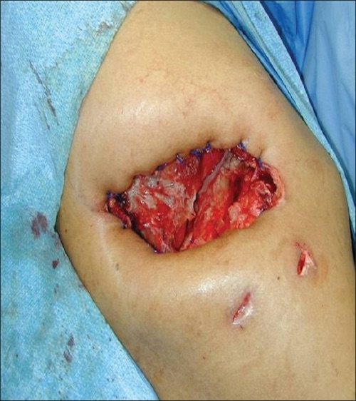
If Not a Surgical Candidate: Eloesser Flap (Open Thoracic Window)
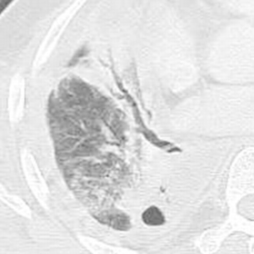
Empyema on CT 1
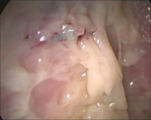
Empyema with Necrotic Purulent Debris on Thoracoscopy 2
Eloesser Flap 3
References
Tsubakimoto M, Murayama S, Iraha R, Kamiya H, Tsuchiya N, Yamashiro T. Can Peripheral Bronchopleural Fistula Demonstrated on Computed Tomography be Treated Conservatively? A Retrospective Analysis. J Comput Assist Tomogr. 2016 Jan-Feb;40(1):86-90. (License: CC BY-NC-ND-4.0)
Patil CB, Dixit R, Gupta R, Gupta N, Indushekar V. Thoracoscopic evaluation of 129 cases having undiagnosed exudative pleural effusions. Lung India. 2016 Sep-Oct;33(5):502-6. (License: CC BY-NC-SA-3.0)
Wait MA, Beckles DL, Paul M, Hotze M, Dimaio MJ. Thoracoscopic management of empyema thoracis. J Minim Access Surg. 2007 Oct;3(4):141-8. (License: CC BY-2.0)