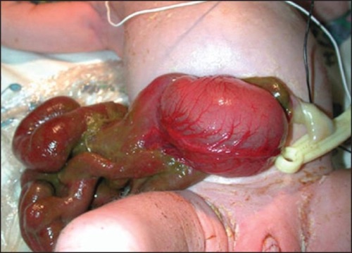
Pediatric Surgery: Abdominal Wall Pathology

Abdominal Wall Defects (Gastroschisis/Omphalocele)

Definitions

  • Omphalocele
    • Larger Defect (> 4 cm) at the Umbilicus
    • Covered by Amniotic Membrane/Peritoneal Sac Mn
      • Covered Bowel is Normal in Appearance
  • Gastroschisis
    • Smaller Defect (< 4 cm), Generally to the Right of the Umbilicus
    • Not Covered by Amniotic Membrane/Peritoneal Sac
      • Uncovered Bowel is Generally Normal at Birth but After 10-20 Minutes will Become Thickened, Matted & Edematous with Fibrous Peel from Amniotic Fluid/Meconium Staining
    • Considered a Surgical Emergency (Bowel Not Covered)
    • Most Common Abdominal Wall Defect

Proposed Pathogenesis

  • Not Entirely Understood
  • Omphalocele:
    • Failure of Lateral Folds to Close
    • Failure of Normal Counterclockwise Rotation Back into Abdomen
  • Gastroschisis:
    • Failure of Umbilical Coelom to Develop
    • Yolk Sac & Vitelline Structures Fail to Incorporate into the Umbilical Cord
    • Intrauterine Umbilical Vein Involution/Rupture

Associations

  • All Have Intestinal Malrotation by Definition
  • Omphalocele:
    • Congenital Anomalies & Midline Defects are More Common
      • Worse Overall Prognosis
    • Cardiac Abnormalities (7-47%)
    • Down Syndrome
    • Cantrell Pentalogy
      • Omphalocele
      • Cardiac Defects
      • Pericardial Defects
      • Cleft Sternum
      • Diaphragmatic Hernia
  • Gastroschisis:
    • Fewer Congenital Anomalies Than Omphalocele
    • Intestinal Atresia More Common

Prenatal Period

  • Most are Found on Prenatal US
  • Cesarean Delivery is Not Necessary & Route of Delivery Does Not Affect Outcomes for Isolated Defects

Treatment

  • Initial Management: Saline-Soaked Gauze Over the Bowel & Resuscitation
    • Gauze Preserves Body Heat, Minimizes Insensible Fluid Loss & Protects Bowel
    • Place NG/OG Tube for Decompression
    • Open Abdominal Wall Defect if Too Tight & Causing Ischemia
  • When Stable: Surgical Repair
    • May Need Silastic Mesh Silo & Delayed Primary Repair if Too Tight
    • Monitor for Abdominal Compartment Syndrome Postoperatively

Omphalocele

Gastroschisis 1

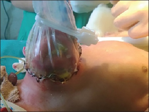
Gastroschisis Silo 2

Umbilical Defects

Umbilical Hernia

Umbilical Cord Hernia

  • Smaller Defect (< 4 cm) at the Umbilicus
  • Covered by Amniotic Membrane/Peritoneal Sac – Often Confused for Omphalocele
    • Not Covered by Skin (Compared to Umbilical Hernia)
  • Only Contains Midgut, Never Liver
    • All Have Malrotation – Although Do Not Typically Cause Obstruction
  • Abdominal Wall Superior to Defect is Normal – Rectus Meets in the Midline at Xiphoid
  • Treatment: Reduction & Primary Fascial Closure

Patent Omphalomesenteric (Vitelline) Duct

  • Presentation:
    • Painless Chronic Mucus Drainage
    • May See Feculent Drainage if the Entire Tract is Patent
    • Small Bowel Obstruction
  • US Shows a Tubular Structure
  • Treatment: Surgical Resection

Omphalitis

  • Definition: Infection of the Umbilicus & Surrounding Tissues
  • Predominately Occurs in the Neonatal Period
  • Most Common in Developing Countries
  • High Mortality: 7-15%
  • Presentation:
    • Start Around 2-3 Days After Birth
    • Umbilical/Periumbilical Pain
    • Umbilical/Periumbilical Erythema & Induration
    • Purulent Drainage from the Umbilical Stump
    • Fever
  • Risk Factors:
    • Intentional Umbilical Nonseverance (“Lotus Birth”) – Umbilical Cord Not Separated After Birth
    • Low Birth Weight
    • Prolonged Labor
    • Prolonged Rupture of Membranes
    • Nonsterile Delivery
    • Maternal Infection
    • Cultural Application of Cow Feces
    • Leukocyte Adhesion Disorders
  • Diagnosis: Clinical
  • Treatment: Broad Spectrum IV Antibiotics
    • Generally Treated for 10 Day Course

Umbilical Granuloma

  • Presentation: Painless Moist Pink-Red Lump of Tissue Seen After Umbilical Cord Has Separated
  • Treatment: Silver Nitrate (Typically Requires Multiple Applications Over Several Weeks)
    • If Fails: Ligation

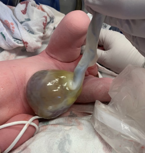
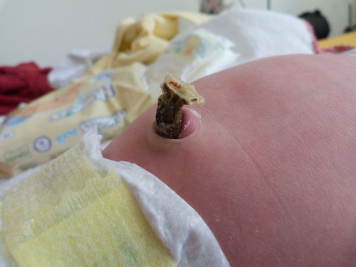
Umbilical Cord Hernia Containing a Meckel’s Diverticulum 3

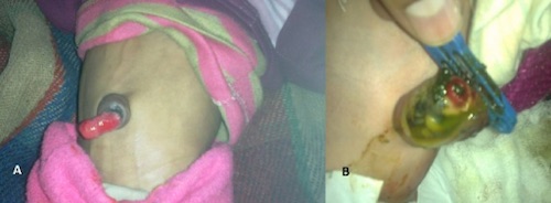
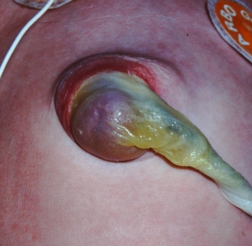
Patent Omphalomesenteric Duct, (A) Prolapsed Patent Duct, (B) Meconium Drainage from Patent Duct 4

Omphalitis 5

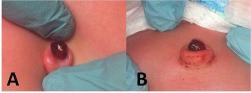
Umbilical Granuloma 6

Mnemonics

Omphalocele vs Gastroschisis

  • Peritoneal Sac Covering:
    • “O” – A Completed Ring Around a Peritoneal Sac
    • “G” – Not a Complete Ring – Not Covered by a Peritoneal Sac
  • Location:
    • “O” at the Belly Button

References

  1. Wright NJ, Zani A, Ade-Ajayi N. Epidemiology, management and outcome of gastroschisis in Sub-Saharan Africa: Results of an international survey. Afr J Paediatr Surg. 2015 Jan-Mar;12(1):1-6. (License: CC BY-NC-SA-3.0)
  2. Dikshit VK, Gupta RK, Gupta AR, Kothari PR, Kamble RS, Kekre GA, Patil PS. Use of composite mesh in gastroschisis: A unique approach. Afr J Paediatr Surg. 2015 Apr-Jun;12(2):148-51. (License: CC BY-NC-SA-3.0)
  3. Gys B, Demaeght D, Hubens G, Ruppert M, Vaneerdeweg W. Herniation of a Meckel’s diverticulum in the Umbilical Cord. J Neonatal Surg. 2014 Oct 20;3(4):52. (License: CC BY-3.0)
  4. Kadian YS, Verma A, Rattan KN, Kajal P. Vitellointestinal Duct Anomalies in Infancy. J Neonatal Surg. 2016 Jul 3;5(3):30. (License: CC BY-3.0)
  5. Stagiryta. Wikimedia Commons. (License: CC BY-SA-4.0)
  6. Alexander G, Walsh R, Nielsen A. Neonatal umbilical mass. West J Emerg Med. 2013 Mar;14(2):163. (License: CC BY-NC-4.0)