Cardiothoracic Surgery: Pneumonia (PNA)

Pneumonia (PNA) – Definitions

Definition

  • Pneumonia (PNA): Acute Infection of Pulmonary Parenchyma

Types

  • Community-Acquired Pneumonia (CAP): Acquired Outside of the Hospital
    • Typical Pneumonia: Typical Organisms Cultured of Standard Media
    • Atypical Pneumonia: Atypical Organisms Not Cultured on Standard Media
  • Healthcare-Associated (Nosocomial) Pneumonia (HCAP): Acquired in the Hospital Setting
    • Hospital-Acquired Pneumonia (HAP): Acquired ≥ 48 Hours After Hospital Admission
      • *Differentiation Between HCAP, HAP & Nosocomial PNA Varies
    • Ventilator-Associated Pneumonia (VAP): Acquired ≥ 48 Hours After Intubation
  • Aspiration Pneumonia: Associated with Aspiration of Material into the Lower Respiratory Tract

Pneumonia (PNA) – Microbiology

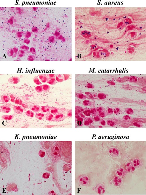
Community-Acquired Pneumonia (CAP) Organisms

  • Typical Organisms:
    • Streptococcus pneumonia – Most Common Cause of CAP
    • Klebsiella pneumonia
    • Hemophilus influenzae
    • Pseudomonas aeruginosa
    • Staphylococcus aureus
  • Atypical Organisms:
    • Legionella
    • Mycoplasma pneumonia
    • Chlamydia pneumonia

Healthcare-Associated Pneumonia (HCAP) Organisms

  • Staphylococcus aureus
    • Methicillin-Sensitive Staphylococcus aureus (MSSA)
    • Methicillin-Resistant Staphylococcus aureus (MRSA) – Most Common Cause of HCAP
  • Pseudomonas aeruginosa
  • Klebsiella pneumonia
  • Escherichia coli
  • Enterobacter spp
  • Acinetobacter spp
  • Streptococcus pneumonia

Common Contaminants

  • Staphylococcus epidermidis
  • Candida spp
  • Streptococcus viridians
  • Corynebacterium diphtheriae

HCAP Multidrug-Resistant (MDR) Risk Factors

  • MDR Risk Factors:
    • IV Antibiotic Use within the Last 90 Days
    • VAP-Specific Risk Factors:
      • Prolonged (≥ 5 Days) Hospitalization Prior to VAP Onset
      • Septic Shock at Time of VAP Onset
      • ARDS Prior to VAP Onset
      • Acute Renal Replacement Therapy Prior to VAP Onset
  • MRSA Risk Factors:
    • Colonization with or Prior Isolation of MRSA
    • ICU/Unit with > 10-20% of S. aureus Isolates Being Methicillin-Resistant
    • ICU/Unit with Unknown MRSA Prevalence
  • MDR Gram-Negative Bacilli (Pseudomonas or Other) Risk Factors:
    • Colonization with or Prior Isolation of MDR Gram-Negative Bacilli (Pseudomonas or Other)
    • HAP-Specific Risk Factors:
      • Structural Lung Disease (Bronchiectasis or Cystic Fibrosis)
      • Respiratory Specimen Gram Stain with Numerous Gram-Negative Bacilli
    • VAP-Specific Risk Factors:
      • ICU with > 10% of Gram-Negative Isolates Resistant to an Agent Considered for Monotherapy
      • ICU with Unknown Local Antimicrobial Susceptibility Rates
  • HAP Risk Factors for Increased Mortality:
    • Requires Ventilator Support for HAP
    • Septic Shock

Pneumonia Gram Stains 1

Pneumonia (PNA) – Diagnosis

Presentation

  • Fever
  • Cough
  • Pleuritic Chest Pain
  • Purulent Sputum
  • Tachypnea
  • Dyspnea
  • Hypoxia
  • Ventilator Changes:
    • Reduced Tidal Volume
    • Increased Inspiratory Pressures
  • Leukocytosis

Diagnosis

  • Gold Standard: Lung Biopsy (Invasive & Rarely Used)
  • Clinical Diagnosis: New Lung Infiltrate on Imaging with Clinical Evidence
    • CXR Has Overall Low Sensitivity & Specificity
  • Culture Supports Diagnosis & Guides Therapy
    • Indicators of Good-Quality:
      • Moderate-Many Polymorphonuclear Leukocytes
      • Few-No Squamous Epithelial Cells
    • VAP Thresholds:
      • Endotracheal Aspirate > 1,000,000 (106) CFU/mL
      • Bronchoalveolar Lavage (BAL) > 10,000 (104) CFU/mL
      • Protected Specimen Brush (PSB) > 1,000 (103) CFU/mL
    • *Should Be Obtained Prior to Initiation of Antibiotics

Culture Types

  • Sputum Culture – Expectorated Sputum, Ideally from a Deep Cough
  • Endotracheal Aspirate – Aspirated Sputum from a Catheter Passed into the Trachea, Possibly Through an Endotracheal Tube
  • Bronchoalveolar Lavage (BAL) – Using a Bronchoscope the Distal Bronchi are Washed with Saline & Then Suctioned for Culture
    • *See Endoscopy: Respiratory Endoscopy
    • Obtains a Large Sample than PSB & Often Preferred
    • Mini-BAL – Catheter Blindly Advanced Through an Endotracheal Tube Until Resistance is Met Followed by Infusion of Three 50 cc Aliquots of Saline & Aspiration
  • Protected Specimen Brush (PSB) – Using a Bronchoscope the Distal Bronchi are Brushed for Culture

Pneumonia on CXR 2

Pneumonia on CT 3

Pneumonia (PNA) – Treatment

CAP Treatment Regimen

  • Primary Treatment: Antibiotics
  • *Antibiotic Regimen Varies Based on Risk Factors, Allergies & Unit of Admission

HCAP Treatment Regimen

  • No MDR Risks: Single-Drug Therapy
  • MRSA Risk: Two-Drug Therapy (Single-Drug Plus MRSA Coverage)
  • MDR Gram-Negative Bacilli Risk: Two-Drug Therapy (Single-Drug Plus MDR Gram-Negative Bacilli Coverage)
  • MDR Risk: Three-Drug Therapy (Single-Drug Plus MRSA Coverage & MDR Gram-Negative Bacilli Coverage)
    • Other Indications for Three-Drug Therapy:
      • HAP with Increased Mortality Risk
      • Both MRSA & Gram-Negative Bacilli Risk

HCAP Antibiotic Options

  • Single-Drugs:
    • Piperacillin-Tazobactam (Zosyn)
    • Cefepime
    • Levofloxacin
    • Meropenem
  • MRSA Coverage:
    • Vancomycin
    • Linezolid
  • MDR Gram-Negative Bacilli Coverage:
    • Aminoglycosides:
      • Amikacin
      • Gentamicin
      • Tobramycin
    • Fluoroquinolones:
      • Levofloxacin
      • Ciprofloxacin
    • Aztreonam

References

  1. Fukuyama H, Yamashiro S, Kinjo K, Tamaki H, Kishaba T. Validation of sputum Gram stain for treatment of community-acquired pneumonia and healthcare-associated pneumonia: a prospective observational study. BMC Infect Dis. 2014 Oct 18;14:534.(License: CC BY-4.0)
  2. Bartziokas K, Daenas C, Preau S, Zygoulis P, Triantaris A, Kerenidi T, Makris D, Gourgoulianis KI, Daniil Z. Vibration response imaging: evaluation of rater agreement in healthy subjects and subjects with pneumonia. BMC Med Imaging. 2010 Mar 11;10:6. (License: CC BY-2.0)
  3. Miyashita N, Sugiu T, Kawai Y, Oda K, Yamaguchi T, Ouchi K, Kobashi Y, Oka M. Radiographic features of Mycoplasma pneumoniae pneumonia: differential diagnosis and performance timing. BMC Med Imaging. 2009 Apr 29;9:7.(License: CC BY-2.0)