Cardiothoracic Surgery: Pulmonary Physiology

Lung Volumes

Lung Volumes

  • Base Volumes:
    • Tidal Volume (TV): Normal Breath
    • Inspiratory Residual Volume (IRV): Maximum Extra Inhaled
    • Expiratory Reserve Volume (ERV): Maximum Extra Exhaled
    • Residual Volume (RV): Volume Unable to Exhale
  • Other Volumes: Mn
    • Inspiratory Capacity (IC) = TV + IRV
    • Functional Residual Capacity (FRC) = ERV + RV
      • Increase: PEEP
      • Decrease: Surgery, ARDS, Trauma
    • Vital Capacity (VC) = TV + IRV + ERV = TLC – RV
      • Aka Functional Vital Capacity (FVC)
    • Total Lung Capacity (TLC) = VC + RV

Factors that Effect

  • Compliance: Volume Produced by a Unit of Pressure Change
  • Elastic Recoil: Ability to Rebound After Inhalation

Muscles of Respiration

  • Inspiration – Elevates Ribs & Sternum
    • Diaphragm (80% of Normal)
    • Accessory Muscles: (20% of Normal)
      • External Intercostals
        • *May Be Considered a Primary Muscle of Inspiration Depending on the Source
      • SCM
      • Scalenes (Anterior, Middle & Posterior)
      • Serratus Anterior & Posterior
      • Levators
  • Expiration – Depresses Ribs & Sternum
    • Mostly Passive Due to Elastic Recoil
    • Forceful Expiration:
      • Internal Intercostals
      • Innermost Intercostals
      • Subcostalis Muscle
      • Abdominal Muscles (Rectus Abdominis, Transverse Abdominis, External Oblique & Internal Oblique)

Intercostal Muscles 1

Disease Changes

  • Obstructive Lung Disease
    • Decreased: Low FEV1 & FEV1/FVC Ratio
      • *FEV1 = Forced Expiratory Volume in 1 Second
    • Increased: TLC & RV
    • Prolonged Expiratory Phase
  • Restrictive Lung Disease
    • Decreased: TLC, RV & FVC

Gas Exchange

Pulmonary Physiology

  • Develops From 7 Months Gestation to 10 Years Old
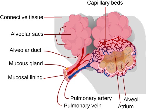
  • Pneumocytes
    • Type I: Gas Exchange
    • Type II: Produce Phosphatidylcholine/Surfactant
      • Lowers Surface Tension & Keeps Alveoli Open
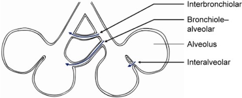
  • Collateral Ventilation:
    • Pores of Kohn: Direct Air Exchange Between Alveoli
    • Channels of Lambert: Air Exchange from Bronchiole to Alveolus
    • Channels of Martin: Air Exchange Between Bronchioles
  • Partial Pressure of Oxygen
    • Highest Point: Pulmonary Capillaries
      • Slightly Less by the Time Blood Reaches the Atrium
    • Lowest Point: Coronary Veins

Ventilation/Perfusion

  • Dead Space: Area Ventilated but Not Perfused
    • Causes Increased PCO2
    • Most Common Cause: Excessive PEEP (Induces Capillary Compression)
  • Shunt: Area Perfused but Not Ventilated
    • Causes Decreased PO2
    • Most Common Cause: Atelectasis

Ventilation/Perfusion (V/Q) Ratio

  • Causes of High V/Q Ratios:
    • Dead Space
    • Normal Lung Apex
    • COPD
    • Pulmonary Embolism
  • Causes of Low V/Q Ratios:
    • Shunting
    • Normal Lung Base
    • Asthma
    • Pulmonary Edema
  • Ratio Changes with Body Positioning

Pulmonary Alveoli 2

Collateral Ventilation 3

Pleural Fluid

Function

  • Serous Fluid
  • Acts as a Lubricant for Parietal & Visceral Pleura to Prevent Adhesion During Respiration

Flow

  • Production: Parietal Circulation (Intercostal Arteries) from Bulk Flow
  • Reabsorption: Lymphatic System
    • Mostly (75%) from Lymphatics of the Parietal Pleura
    • Visceral Pleural Plays No Significant Role in Drainage

Volume

  • Normal Amount Present: 10-20 cc
  • Fluid Turnover Ability: 1-2 L/Day
    • Can Increase Up to 40x Normal Rates
    • *Requires a Profound Increase in Production or Blockage of Lymphatics to Initiate Fluid Accumulation

Mnemonics

Capacities vs Residual Volumes

  • C-C: Capacity = Combination of Others

References

  1. CF CF. Wikimedia Commons. (License: CC BY-SA-4.0)
  2. Lady of Hats. Wikimedia Commons. (License: Public Domain)
  3. Koster TD, Slebos DJ. The fissure: interlobar collateral ventilation and implications for endoscopic therapy in emphysema. Int J Chron Obstruct Pulmon Dis. 2016 Apr 13;11:765-73.(License: CC BY-NC-3.0)