Ia: ≤ 4.0 cm & Confined to Kidney
Ib: > 4.0 cm, ≤ 7.0 cm & Confined to Kidney
LN+
Mets+
II
IIa: > 7.0 cm, ≤ 10.0 cm & Confined to Kidney
IIb: > 10.0 cm & Confined to Kidney
III
IIIa: Extends into Renal Vein or its Segmental Branches, Invades Pelvicalyceal System or Invades Perirenal and/or Renal Sinus Fat but Not Beyond Gerota’s Fascia
IIIb: Grossly Extends into the Vena Cava Below the Diaphragm
IIIc: Grossly Extends into the Vena Cava Above the Diaphragm or Invades the Wall of the Vena Cava
IV
Invades Beyond Gerota’s Fascia
Stage
T
N
M
I
T1
N0
M0
II
T2
N0
M0
III
T3
N0
M0
T1-3
N1
M0
IV
T4
Any N
M0
Any T
Any N
M1
Diagnosis
Primary Evaluation: CT or MRI
> 15-20 Hounsfield Units (HU) is Indicative of RCC
Unable to Reliably Diagnose RCC in Solitary Small Lesions – Generally Requires Resection
Renal Mass Biopsy (RMB) May Be Considered in Select Circumstances
Biopsy Often Avoided Due to Associated Risks:
High Nondiagnostic Rate
Occasional False-Negatives
Potential for Cystic Tumor Spillage
Treatment
Primary Treatment: Surgical Resection
Intermediate-High Risk Patients Should Be Offered Adjuvant Pembrolizumab (PD-1 Inhibitor)
Locally Advanced RCC (Invading IVC) is Still Primarily a Surgical Disease
Metastatic Disease:
Nephrectomy with Mastectomy May Be Considered for Limited Metastatic Disease
Factors Associated with Improved Outcomes After Metastasectomy:
Complete Resection
Solitary Metastatic Lesions
Age < 60 Years
Small Tumor Size
Pulmonary Metastasis
Metachronous Metastatic Disease
May Consider Cytoreductive Nephrectomy Alone in Select Patients
Extent of Surgical Resection
Indications for Partial Nephrectomy:
Small T1a Lesions (< 4 cm)
Solitary Kidney or Abnormal Contralateral Kidney
Bilateral Tumors
Preexisting Chronic Kidney Disease (CKD) or Proteinuria
Multifocal Masses
Other Comorbidities May Impact Future Renal Function
Known Familial RCC
Indications for Radical Nephrectomy:
Central Location in Kidney
Suspected Lymph Node Disease
Extension into Renal Vein, IVC or Adrenal Gland
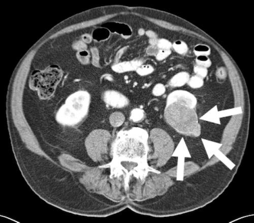
Renal Cell Carcinoma on CT 2
References
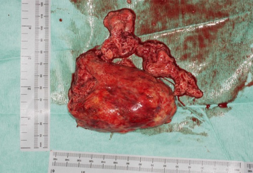
Parissis H, Akbar MT, Tolan M, Young V. Surgical resection of a renal cell carcinoma involving the inferior vena cava: the role of the cardiothoracic surgeon. J Cardiothorac Surg. 2010 Nov 5;5:103. (License: CC BY-2.0)
Nguyen BD, Roarke MC. Renal Cell Carcinoma with Paraneoplastic Manifestations: Imaging with CT and F-18 FDG PET/CT. Radiol Case Rep. 2015 Dec 7;2(3):72. (License: CC BY-NC-ND-4.0)