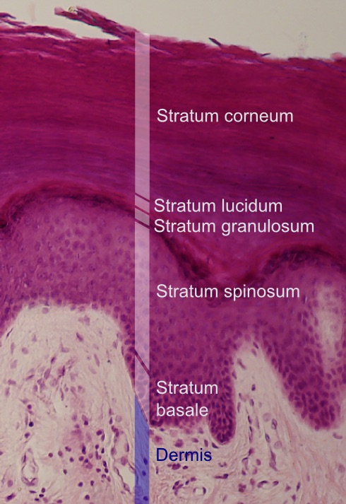
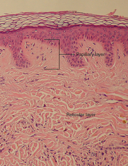
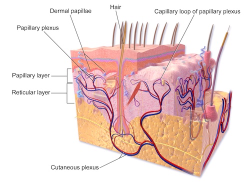
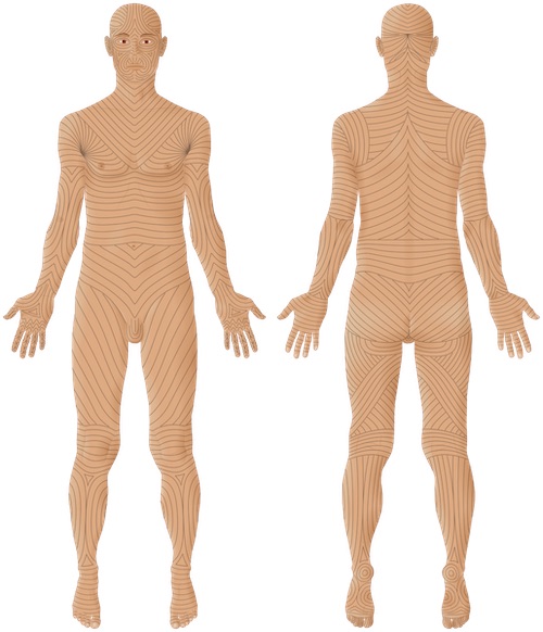
Skin & Soft Tissue: Anatomy & Physiology Anatomy Skin LayersEpidermisDermisSubcutaneous Fascia (Hypodermis)EpidermisLayers: Mn Stratum Corneum – Thickest & Outermost LayerStratum Lucidum – Present Only in Thicker Skin (Palms & Soles)Stratum GranulosumStratum SpinosumStratum Basale (Stratum Germinativum)Basement MembraneCell Types:KeratinocytesPrimary Cell Type of the EpidermisOriginate in the Basal LayerProduce Keratin & Secrete Lipids to Form a Water BarrierMelanocytesDerived from Neural Crest CellsProduce Melanin & Transfer it to KeratinocytesSkin Tone is Based on Melanin ProductionMelanocyte Density is Generally SimilarLangerhans CellsDendritic Cells (Antigen Presenting Cells)Merkel CellsSensory Receptors for Light TouchHighest Density in FingertipsDermisLayers:Papillary Layer – Outermost Layer Just Deep to the Basement MembraneMade of Loose Connective Tissue with Loosely-Packed CollagenDermal Papillae – Extensions into the Epidermis that Form Papillary Ridges (Fingerprints)Reticular Layer – Deeper LayerMade of Dense Connective Tissue with Densely-Packed Collagen (Provides StrengthTensile Strength Provided by Type I CollagenSensory Nerves:Pacinian Corpuscle: Sense Deep PressureMeissner Corpuscle: Tactile/Low-Frequency StimulationRuffini Corpuscle/Ending: Sense Sustained Pressure & WarmthKrause End-Bulbs: Sense ColdSweat Glands:Eccrine Sweat GlandsDistribution: Everywhere but Highest in Palms/SolesSecretion: Aqueous Fluid for CoolingSecrete Directly onto the SkinApocrine Sweat GlandsDistribution: Axilla, Groin, Gluteal & Inframammary FoldsSecretion: Cloudy Thick Sweat Mn Secrete into the Pilary Canal of Hair FolliclesInactive Prior to PubertyLanger’s Lines – Topological Cleavage Lines that Define TensionCorrespond to Collagen Alignment in the Reticular DermisOften Used in Making Surgical IncisionsSubcutaneous Tissue (Hypodermis)Primarily Used for Fat Storage (Adipocytes)Also Contains Nerves, Blood Vessels & Lymphatics Layers of the Epidermis 1 Layers of the Dermis 2 Contents of the Dermis 3 Langer’s Lines 4 Mnemonics Layers of the Epidermis“Come, Let’s Get Sun Burned”CorneumLucidumGranulosumSpinosumBasaleApocrine vs Eccrine Sweat GlandsDirty Apes (APE-ocrine) Stink References Haggstrom M. Wikimedia Commons. (License: CC BY-SA-3.0)OpenStax College. Wikimedia Commons. (License: CC BY-3.0)Blaus B. Wikimedia Commons. (License: CC BY-3.0)Goran Tek-en. Wikimedia Commons. (License: CC BY-SA-4.0)