Small Intestine: Small Intestine Diverticula

Small Intestine Diverticulosis

Description

  • False Diverticulum
  • Herniate on the Mesenteric Border Near Arterial Entrance

Locations

  • Duodenum – Most Common (45-80%)
    • Most Common at 2nd Portion Near Ampulla
    • Usually Solitary
  • Jejunum – Second Most Common
    • Commonly Multiple
  • Ileum
    • Commonly Multiple

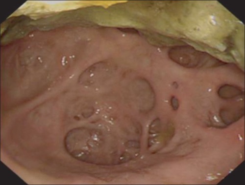
Duodenum Diverticulum on EGD 1

Presentation

Treatment

  • ASx: Observe
  • Sx: Diverticulectomy
    • If Second Portion of the Duodenum with Cholangitis/Pancreatitis: ERCP with Sphincterotomy/Stent & Diverticulectomy When Resolved
    • Consider Prophylactic Cholecystectomy for Duodenal Diverticula
    • Consider Segmental Resection of Jejunoileal Diverticula if Necessary

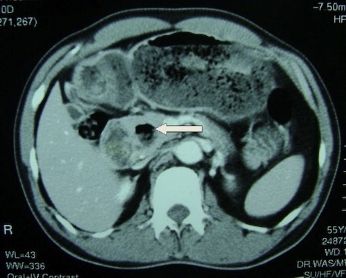
Duodenum Diverticulum on CT 2

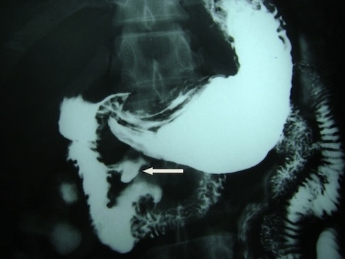
Duodenum Diverticulum on UGI 2

Meckel’s Diverticulum

Pathology

  • Remnant of a Persistent Vitelline Duct Due to Failure of Obliteration
  • The Most Common Congenital Small Bowel Abnormality
  • Most Common Cause of Painless GI Bleed in Peds
  • Most Common Neoplasia: Carcinoid

Description

  • True Diverticulum
  • Herniate on the Antimesenteric Border

Heterotopic Tissue

  • Gastric Tissue (Most Common – 75%)
    • Most Likely to Produce Sx
    • Causes Bleeding
  • Pancreatic Tissue (15%)
    • Causes Inflammation

Presentation

  • Bleeding – Most Common Presentation
    • From Ulceration on the Mesenteric Border
    • Ulcer Caused by Acid Produced by Ectopic Gastric Tissue
  • Obstruction
    • Causes: Intussusception, Volvulus, Torsion, Littre’s Hernia
  • Meckel’s Diverticulitis
    • Inflammation from Luminal Obstruction
    • Causes: Foreign Body, Enterolith, Inflammatory Tissue or Tumor

Rules of 2’s

  • 2 Feet from Ileocecal Valve
  • 2 Inches Long
  • 2 cm Diameter
  • 2% of the Population
  • 2x as Common in Males
  • Most Often Presents by 2 Years Old, But Can Be Seen in Adults
  • 2% Become Symptomatic
  • 2 Presentations (Bleeding or Obstruction)
  • 2 Types of Tissue

Diagnosis

  • Meckel’s Scintigraphy Scan (99Tc)
    • Detects Gastric Mucosa
    • Does Not Detect Pancreatic Tissue or Hemorrhage
    • 85% Sensitive in Pediatric Patients
      • Much Less Sensitive in Adults (Due to Atrophic Gastric Mucosa)

Treatment

  • Incidental
    • On Imaging: Observation
    • Intraoperative: Observation
      • Possible Indication for Surgery if Found Incidentally:
        • Length > 2 cm
        • Palpable Abnormality
        • Fibrous Bands
      • Surgical Options:
        • Diverticulectomy Generally Preferred
        • Consider Segmental Resection if Broad Base (> 2 cm) or Palpable Abnormality at the Base
      • *Historically Diverticulectomy was Dogma but Newer Evidence Recommends Observation for Most Incidental Meckel’s
  • Diverticulitis
    • Uncomplicated: Diverticulectomy
    • Complicated: Segmental Resection
      • Indications:
        • Perforated
        • Base Inflamed
        • Wide Base (> 1/3 Diameter of Bowel or Neck > 2 cm)
  • Bleeding: Diverticulectomy vs Segmental Resection
    • Reason for Segmental Resection: Ulcer on Mesenteric Border & Occasionally Distal to the Antimesenteric Diverticulum
    • *Historically Segmental Resection was Dogma but Newer Evidence Questions Diverticulectomy for Bleeding Meckel’s with Rare Incidence of Repeat Bleeding

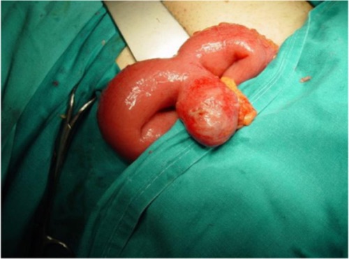
Meckel Diverticulum 3
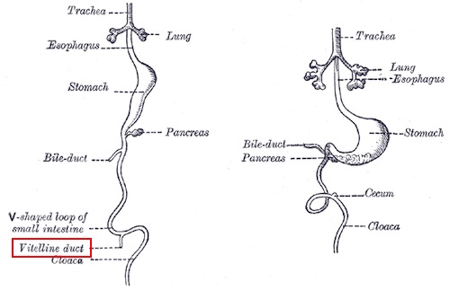
Vitelline Duct 4

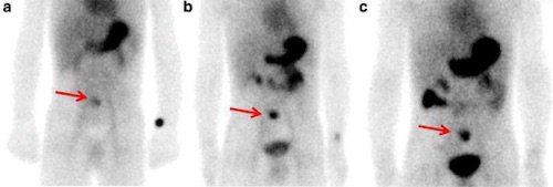
Meckel’s on Scintigraphy 5

References

  1. Kim PS, Jung EJ, Bang HY. Small bowel diverticulosis in patient with early gastric cancer. Ann Surg Treat Res. 2014 Oct;87(4):209-12. (License: CC BY-NC-3.0)
  2. Seneviratne SA, Samarasekera DN. Massive gastrointestinal haemorrhage from a duodenal diverticulum: a case report. Cases J. 2009 Jul 2;2:6710. (License: CC BY-2.0)
  3. Dimitriou I, Evaggelou N, Tavaki E, Chatzitheoklytos E. Perforation of Meckel’s diverticulum by a fish bone presenting as acute appendicitis: a case report. J Med Case Rep. 2013 Oct 2;7:231. (License: CC BY-2.0)
  4. Gray H. Anatomy of the Human Body (1918). Public Domain.
  5. Xie Q, Ma Q, Ji B, Gao S, Wen Q. Incremental value of SPECT/CT in detection of Meckel’s diverticulum in a 10-year-old child. Springerplus. 2016 Aug 5;5(1):1270. (License: CC BY-4.0)