Endocrine: Thyroid Nodule Management

Thyroid Nodule Management

Thyroid Nodules

  • High-Prevalence in General Population: 37-57%
  • Majority (90%) are Benign
    • Only 50% in Peds
  • Carcinoma Risk Factors:
    • Age Extremes (Pediatrics & Elderly)
    • Male Sex
    • History of Radiation to Head & Neck
      • Most Likely Papillary if From a History of XRT
    • Solitary Nodule (vs Multinodular)
    • Large ≥ 2 cm

Initial Management

  • Initial Testing: Thyroid Function Tests (TFTs) & Ultrasound (US)
  • If Patient Is Hyperthyroid May First Consider Scintigraphy (Radioiodine Uptake Scan)
    • Hot Nodule/Hyperfunctioning: Benign – No Further Testing
    • Cold Nodule: Risk for Malignancy – Further Testing/US is Indicated

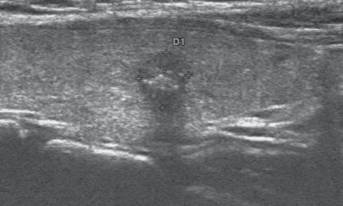
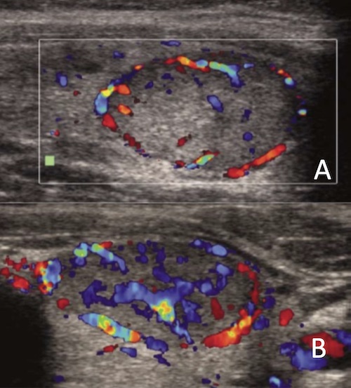
Ultrasound (US) – Concerning Features

  • Most Specific:
    • Taller > Wide (Normal Grows Radially)
    • Microcalcifications
  • Heterogenous
  • Hypoechoic
  • Solid (vs Cystic)
  • Lobulated/Irregular Margins
  • Hypervascular

Indications for Fine Needle Aspiration/FNA (Based on US)

  • Intermediate-High Suspicion: ≥ 1.0 cm
  • Low Suspicion: ≥ 1.5 cm
  • Very-Low Suspicion: ≥ 2.0 cm
  • Purely Cystic: FNA Not Indicated

Bethesda System for Reporting Thyroid Cytopathology (Based on FNA)

  • Category I: Nondiagnostic or Unsatisfactory
  • Category II: Benign
  • Category III: Undetermined Significance
    • Atypia of Undetermined Significance (AUS)
    • Follicular Lesion of Undetermined Significance (FLUS)
  • Category IV: Follicular Neoplasm or Suspicious for a Follicular Neoplasm
  • Category V: Suspicious for Malignancy
  • Category VI: Malignant 

Thyroid Nodule Malignant Findings: Solid, Heterogenous, Hypoechoic, Microcalcifications 1

Thyroid Nodule Vascularity; (A) Normal/Benign, (B) Hypervascular/Malignant 2

Risk of Malignancy

Category Risk if NIFTP ≠ CA Risk if NIFTP = CA
I 5-10% 5-10%
II 0-3% 0-3%
III 6-18% 10-30%
IV 10-40% 25-40%
V 45-60% 50-75%
VI 94-96% 97-99%
    • NIFTP = Noninvasive Follicular Thyroid Neoplasm with Papillary-Like Nuclear Features
    • CA = Carcinoma

Management (Based on Bethesda Category)

  • Category I: Repeat FNA
    • Consider Excision if Repeat FNA Again Results Category I
  • Category II: Clinical Follow Up
  • Category III: Repeat FNA (May Consider Molecular Testing)
  • Category IV: Lobectomy/Hemithyroidectomy (May Consider Molecular Testing)
  • Category V: Lobectomy/Hemithyroidectomy vs. Total Thyroidectomy
  • Category VI: Lobectomy/Hemithyroidectomy vs. Total Thyroidectomy

Molecular Testing (Afirma, ThyroSeq, etc.)

  • Recent Advancement that Has Reduced the Need for Unnecessary Surgery
  • Tests for High-Risk Mutations (RET, BRAF, VEGFR, etc.)
  • Better Used to Rule-Out Malignancy than to Rule-In
    • Negative Predictive Value (NPV): > 95%
    • Positive Predictive Value (PPV): 40-80%
    • Sensitivity: 90%
    • Specificity: 50-90%

References

  1. Chen M, Zhang KQ, Xu YF, Zhang SM, Cao Y, Sun WQ. Shear wave elastography and contrast-enhanced ultrasonography in the diagnosis of thyroid malignant nodules. Mol Clin Oncol. 2016 Dec;5(6):724-730. (License: CC BY-NC-ND-4.0)
  2. Salehi M, Nalaini F, Izadi B, Setayeshi K, Rezaei M, Naseri SN. Gray-scale vs. color doppler ultrasound in cold thyroid nodules. Glob J Health Sci. 2014 Nov 26;7(3):147-52. (License: CC BY-3.0)