Trauma: Traumatic Brain Injury (TBI)

Traumatic Brain Injury (TBI)

General

  • Causes:
    • Most Common Leading to Hospitalization: MVC
    • Most Common Leading to Death: GSW
    • Most Common in Elderly: Falls
  • Classification:
    • Mild: GCS 13-15
    • Moderate: GCS 9-12
    • Severe: GCS ≤ 8
  • Coagulopathy Develops from Tissue Thromboplastin Release

Epidural Hematoma (EDH)

  • Bleeding Between the Dura Mater & Skull
  • Most Common Source: Middle Meningeal Artery
    • Associated with Temporal Fractures
  • CT Appearance: Lentiform/Biconvex, Does Not Cross Midline
  • “Lucid Interval” – A Relatively Normal Period of Time from the Initial Concussion to Subsequent Coma

Subdural Hematoma (SDH)

  • Bleeding Between the Dura Mater & Arachnoid Mater
  • Most Common Intracranial Hemorrhage
  • Source: Venous Plexus/Bridging Veins
    • Acceleration/Deceleration Injury
  • CT Appearance: Crescent, Crosses Midline
  • Can Be Chronic in Elderly After Falls

EDH 1

SDH 2

Subarachnoid Hemorrhage (SAH)

SAH 3

Xanthochromia 4

Diffuse Axonal Injury (DAI)

  • A Diffuse Shearing Injury of Brain Axons
  • Most Common Cause: Rotational Force of Acceleration/Deceleration Impact
  • May Not Be Apparent on CT
  • MRI Appearance:
    • Punctate Hemorrhages
    • Blurring of Grey-White Interface
  • Very Poor Prognosis

DAI on MRI 5

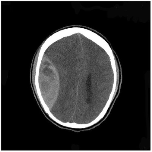
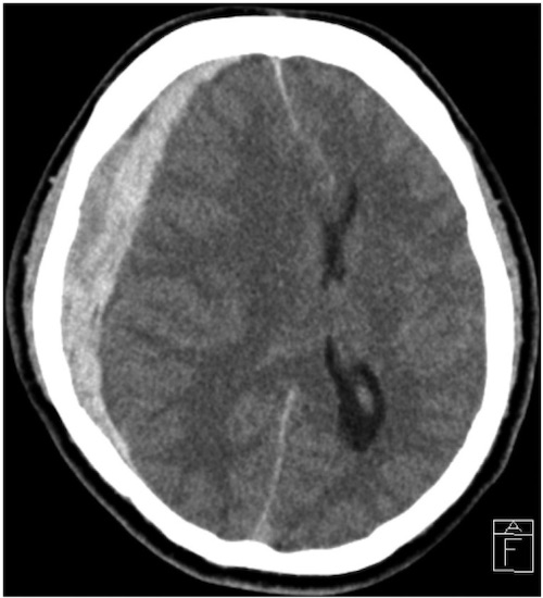
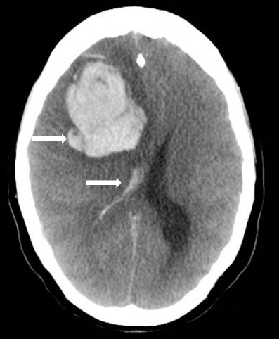
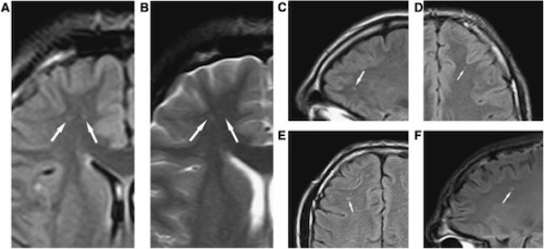
Cerebral Contusion (Parenchymal/Hemorrhagic Contusion)

  • Bruising of the Brain from Multiple Punctate Hemorrhages
    • May See Surrounding Edema & Necrosis
  • Most Remain Small & Surgically Insignificant
  • Evolve Over Time – May Worsen or Not Even Be Evident on Initial CT
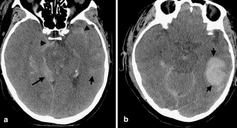
  • Often See “Coup & Countercoup” Injuries
    • “Coup” – Injury at the Site of Head Impact
    • “Countercoup” – Injury Remote from the Site of Head Impact (Classically Directly Opposite)
  • Most Common Sites: Frontal Base and Anterior Temporal Lobes

Cerebral Contusion 6

Secondary Brain Injury

  • Injury That Develops After the Initial/Primary Brain Injury from Metabolic & Physiologic Derangements
  • Risk Factors:
    • Hypotension
      • Reduced CPP Causes Ischemia
      • Autoregulation with Arteriole Vasodilation Causes Increased ICP
      • Doubles the Mortality
    • Hypoxia

Treatment

  • Admit to ICU if Moderate-Severe Injury
  • Seizure Prophylaxis:
    • Indications: Severe TBI (Not Mild-Moderate)
    • 1 Week of Levetiracetam (Keppra) or Fosphenytoin
    • Reduce Risk of Early Seizures
    • No Reduced Risk for Late Seizures or Post-Traumatic Epilepsy
  • Surgical Decompression Indications:
    • Epidural Hematoma
      • > 30 cc Volume
      • GCS ≤ 8 & Pupil Abnormality
    • Subdural Hematoma
      • > 10 mm Thick
      • > 5 mm Midline Shift
      • GCS ≤ 8 & Pupil Abnormality or GCS Decreased by ≥ 2
    • Subarachnoid Hemorrhage
      • Posterior Fossa Causing Mass Effects
      • Cerebral Hemispheres > 50 cc
        • Or > 20 cc with Midline Shift > 5 mm & GCS ≤ 8
  • Anticoagulation Management:
    • If on Coumadin: Repeat CT for Possible Delayed Bleed
    • Start Prophylactic Anticoagulation Within 24-48 Hours of a Stable Head CT
  • *Use of Beta-Blockers to Blunt Sympathetic Activation Cascade After TBI is Evolving

Brain Injury Guidelines (BIG)

Variables BIG 1 BIG 2 BIG 3
LOC Yes/No Yes/No Yes/No
Neuro Exam Normal Normal Abnormal
Intoxication No No/Yes No/Yes
CAP (Coumadin, Aspirin, Plavix) No No Yes
Skull Fracture No Non-displaced Displaced
SDH < 4 mm 5-7 mm > 8 mm
EDH < 4 mm 5-7 mm > 8 mm
IPH (Locations) < 4 mm (x1) 5-7 mm (x2) > 8 mm (Multiple)
SAH Trace Localized Scattered
IVH No No Yes
  • Management:
    • BIG 1: Observe for 6 Hours
    • BIG 2: Admit & Observe for 24 Hours
    • BIG 3: Admit, Repeat Head CT & Consult Neurosurgery

Intracranial Pressure (ICP)

Monro-Kellie Doctrine

  • The Sum of the Volumes of Brain Matter, CSF & Intracranial Blood is Constant
    • An Increase in One Will Cause a Decrease in One or Both of the Others
  • Therefore an Elevated ICP will Result in Decreased Cerebral Perfusion Pressure & Risk for Herniation

Values

  • Intracranial Pressure (ICP)
    • Normal: 10 mmHg
    • Goal: < 20 mmHg
    • Peaks 48-72 Hours After Trauma
  • Cerebral Prefusion Pressure (CPP)
    • CPP = MAPICP

Signs of Elevation

  • Cushing’s Triad
    • From Increased ICP
      • Causes Increased Sympathetic & Parasympathetic Activity
      • Late Sign – Suggests Impending Herniation
    • Triad:
      • HTN (Widened Pulse Pressure)
      • Bradycardia
      • Irregular Breathing Pattern
    • Stages:
      • First Stage
        • From Increased ICP
        • Increases Sympathetic Activity
        • Increases BP & HR
      • Second Stage
        • From Aortic Arch Baroreceptors Due to HTN
        • Increases Parasympathetic Activity
        • Decreases HR
      • Third Stage
        • From High ICP, HR Changes & Endogenous Stimuli
        • Distorts Brainstem Pressures
        • Irregular Breathing
  • Uncal Herniation
    • Earliest Sign: Eyes (Ptosis, Anisocoria (Unequal Pupil Size) & Impaired Movement)
    • Dilated/Blown Pupil
      • Indicates Pressure on Ipsilateral Oculomotor CN III
  • Pain
  • Double Vision (Due to CN VI Palsy)

ICP Monitors

  • Indications:
    • Abnormal CT & GCS ≤ 8
    • Normal CT & ≥ 2 of:
      • Age > 40
      • Motor Posturing
      • SBP < 90
  • Types:
    • Bolt (Codman ICP Monitor)
      • Bolt Placed into the Parenchyma with Fiberoptic Monitoring of ICP
      • Only Measures ICP
    • External Ventricular Drain (EVD/Ventriculostomy)
      • Drain Placed into the Ventricle
      • Measures ICP & Allows Therapeutic CSF Drainage

CPP Management

  • Goal CPP > 60 mmHg
    • Range 50-70 mmHg
    • Goal: Avoid Secondary Brain Injury
  • Initial Measures:
    • IVF (NS)
    • Pressor Support – Avoid Hypotension
    • Elevate Head-of-Bed to 30 Degrees – Allow Adequate Venous Drainage
    • Sedation & Analgesia – Decrease Pain & Metabolic Demand
    • Intermittent CSF Drainage
    • Normothermia – Fever Increase Metabolic Demand & Blood Flow
    • Avoid LP (Precipitates Herniation)
  • Secondary Options:
    • Osmotic Therapy (Goal Na 145-155)
      • Hypertonic Saline or Mannitol Boluses (Not Continuous)
        • Avoid Mannitol if Hypotensive or Hypovolemic
    • Hyperventilate (PaCO2 30-35) – Induce Cerebral Vasoconstriction
    • Exchange Parenchymal Pressure Monitor to External Ventricular Drain
  • Refractory Treatment:
    • Decompressive Craniectomy
      • Improved Survival but More Survivors are Dependent on Others and Higher Risk of Unfavorable Outcomes (RESCUEicp and DECRA Trials)
    • Neuromuscular Paralysis
    • Barbiturate Coma
    • Hypothermia

References

  1. Yogarajah M, Agu CC, Sivasambu B, Mittler MA. HbSC Disease and Spontaneous Epidural Hematoma with Kernohan’s Notch Phenomena. Case Rep Hematol. 2015;2015:470873. (License: CC BY-3.0)
  2. Choi HJ, Lee JI, Nam KH, Ko JK. Acute Spontaneous Subdural Hematoma due to Rupture of a Tiny Cortical Arteriovenous Malformation. J Korean Neurosurg Soc. 2015 Dec;58(6):547-9. (License: CC BY-NC-3.0)
  3. Al-Hameed FM. Spontaneous intracranial hemorrhage in a patient with Middle East respiratory syndrome corona virus. Saudi Med J. 2017 Feb;38(2):196-200. (License: CC BY-NC-SA-3.0)
  4. Kwon SK, Kim MW. Pseudo-Froin’s syndrome, xanthochromia with high protein level of cerebrospinal fluid. Korean J Anesthesiol. 2014 Dec;67(Suppl):S58-9. (License: CC BY-NC-3.0)
  5. Gandy S, Ikonomovic MD, Mitsis E, Elder G, Ahlers ST, Barth J, Stone JR, DeKosky ST. Chronic traumatic encephalopathy: clinical-biomarker correlations and current concepts in pathogenesis. Mol Neurodegener. 2014 Sep 17;9:37. (License: CC BY-2.0)
  6. Kim JJ, Gean AD. Imaging for the diagnosis and management of traumatic brain injury. Neurotherapeutics. 2011 Jan;8(1):39-53. (License: CC BY-NC-2.0)