Abdominal Wall: Ventral Hernias

Typical Ventral Hernias

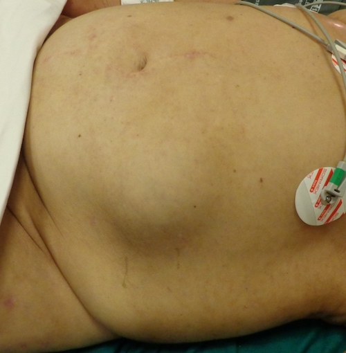
Ventral Hernia Definition

  • Definition: Hernia of the Anterior Abdominal Wall

Types

  • Incisional Hernia
    • Hernia Through a Prior Incision
    • Port-Site Hernia – Incisional Hernia Through a Prior Port Site
    • Develop in 10-15% of Incisions
      • Only a 0.0-0.5% Risk in Pfannenstiel Incisions
    • Risk Factors:
      • Surgical Site Infection (Highest)
      • Obesity
      • Diabetes
      • Increased Intraabdominal Pressure (COPD, Cystic Fibrosis, etc.)
      • Malnutrition
      • Immunosuppression
      • Midline Incisions (Less Common with Transverse or Oblique Incisions)
      • Vertical Incisions
      • Use of Bladed Trocars
    • Efforts to Avoid Port-Site Hernias:
      • Close All Ports ≥ 10 mm
        • Consider Closing 5 mm Under Excessive Traction or in Children
      • Include All Layers in Closure
      • Use the Fewest Ports Possible with the Smallest Diameter
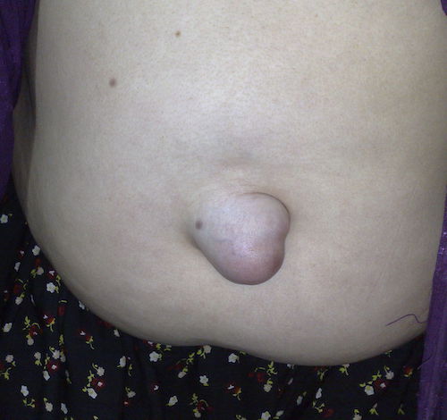
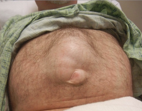
  • Umbilical Hernia
    • Hernia Through the Umbilical Ring
    • 3x More Common in Women
      • 90% of Women Develop an Umbilical Hernia During Pregnancy
    • Strangulation More Common in Men
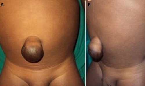
    • Proboscoid (Elephant-Trunk) Hernia
      • Definition: Large Umbilical Hernia with Excessive Stretching of the Skin Resembling a Trunk
      • Named After the Nose of a Proboscis Monkey
      • Usually At Least a Few cm in Diameter
  • Epigastric Hernia
    • Hernia Between the Umbilicus & Xiphoid Along the Linea Alba
    • Usually Small Containing Preperitoneal Fat
    • More Common Above the Umbilicus Than Below
      • Obliterated Umbilical Vessels & Urachus Reinforce Abdominal Wall Below
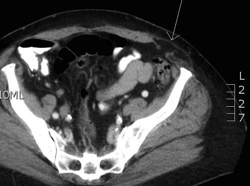
Diagnosis

  • Generally a Clinical Diagnosis
  • US is the Preferred Imaging Modality If Uncertain
    • Can Also Evaluate for Multiple Defects if Diagnosis is Uncertain
    • More Cost Effective than CT & Allows Dynamic Assessment with/without Valsalva
  • Consider CT for Evaluation of Large/Complex Defects

Treatment

  • Small & ASx: Observe
  • Large or Sx: Repair
  • Mesh Indications: ≥ 1-2 cm
    • *In General, if a Mesh Can Fit Through It Should be Used
  • Component Separation
    • Indications:
      • Multiple Defects Unable to Close with Mesh
      • Large Defect Unable to Close Primarily
      • Large Recurrence that Failed Suture Closure
      • Giant Omphalocele
    • Relative Contraindications:
      • Extensive Destruction of Abdominal Wall Components
      • Compromise of Epigastric Arterial Supply (DIEP Flap, etc.)
      • Active Infection
  • Emergent or Contaminated (Necrotic Bowel) Options:
    • Suture Repair
    • Mesh Repair – May Consider Absorbable Mesh but Never Use Nonabsorbable Mesh in Contaminated Cases
    • Staged Repair
      • Close Skin with Planned Ventral Hernia & Delayed Definitive Repair
      • Some Consider the Best Chance for a Good Repair

Open vs Minimally Invasive Comparison

  • Open Surgery:
    • Preferred for Large Defects (> 10 cm)
    • Preferred for Loss of Abdominal Domain
    • Preferred if Bowel is Compromised with Necessary Resection
      • Incarceration Alone Can Be Done Laparoscopically
  • Minimally Invasive:
    • Decreased Risk of Wound Complications – Particularly in Obese
      • Includes Hematoma, Seroma & Surgical Site Infection
    • Shorter Length of Stay
    • Comparable Operative Time
    • Provides Better Visualization of Multiple Defects (Avoids Larger-Than-Needed Incisions)
  • Equivalent Recurrence Rates

Umbilical Hernia 1

Proboscoid Hernia 2

Spigelian (“Semilunar”) Hernia

Basics

  • Definition: Hernia Along the Spigelian Line (Aponeurotic Band at Lateral Border of the Rectus Abdominis)
  • Most are Interparietal – Herniates Between External & Internal Obliques (Can Be Confused with Rectus Sheath Hematoma)
  • Tend to Have Small/Narrow Neck (1-2 cm)
    • High Risk of Incarceration/Strangulation

Location

  • Most Common at the Junction of the Spigelian Line & the Arcuate Line
  • “Spigelian Hernia Belt” – Area Between the Horizontal Lines from Umbilicus to the ASIS (About 6 Inches)
    • Due to Variable Position of the Arcuate Line
    • Contains 90% of Spigelian Hernias

Diagnosis

  • Dx: CT (Often Difficult to Palpate)

Treatment

  • Tx: Repair All

Spigelian Hernia on CT 3

Spigelian Hernia 4

Other Ventral Hernias

Arcuate Line Hernia

  • Hernia Under the Arcuate Line
    • Preperitoneal Fold Ascending Upward Between the Posterior Aponeurotic Sheath & Rectus Abdominis Muscle
    • Can Be Unilateral or Bilateral
  • Defined as an Internal Hernia – No Defect Through the Abdominal Wall
  • Majority are ASx with a Wide Hernia Orifice
  • Often Misdiagnosed as a Spigelian Hernia

Posterior Rectus Sheath Hernia

  • Hernia Through the Posterior Rectus Sheath
  • Forms a Mass Within the Rectus Muscle
  • Many are Interparietal

Diastasis Recti

  • Definition: Separation of Rectus Abdominis Pillars
    • Fascia Intact (Not a True Hernia)
  • Most Common Postpartum or After Weight Loss
  • Presentation:
    • Many are Asymptomatic
    • Can Cause Abdominal Pain
    • Prominent Ridge Extending from the Xiphoid to Umbilicus
    • Midline Fusiform Bulge Rises from Increased Intraabdominal Pressure (Sit-Up)
  • Diagnosis: Clinical Examination
    • US (Preferred) or CT Can Be Used if Diagnosis Uncertain
  • Tx: Wall Strengthening & Weight Loss
    • If Large/Significant Sx: Surgical Rectus Sheath Plication
      • Add Abdominoplasty if Excess Skin
      • Can Improve Pulmonary & Abdominal Wall Function

Diastasis Recti 5

References

  1. Wikimedia Commons (License: CC 1-1.0)
  2. Ashu EE, Leroy GM, Aristide BG, Joss BM, Bonaventure J, Patrick SE, Myriam FG. Double half-cone flap umbilicoplasty for proboscoid umbilical hernia in a 2 years old child with satisfactory results 2 years later. Pan Afr Med J. 2015 Sep 17;22:44. (License: CC BY-2.0)
  3. Wikimedia Commons (License: Public Domain)
  4. Wikimedia Commons (License: CC BY-SA-3.0)
  5. Cheesborough JE, Dumanian GA. Simultaneous prosthetic mesh abdominal wall reconstruction with abdominoplasty for ventral hernia and severe rectus diastasis repairs. Plast Reconstr Surg. 2015 Jan;135(1):268-276. (License: CC BY-NC-ND-3.0)