Anorectal: Abscess & Fistula

Anorectal/Perirectal Abscess

Basics

  • Abscess of the Anorectal Region
  • Most Common Origin: Anal Glands
  • 4-31% Will Recur
  • 33-50% Will Develop Fistula-In-Ano

Causes

  • Cryptoglandular Disease – Most Common
    • Anal Glands/Crypts of Morgagni at the Dentate Line are Blocked Causing Infection
  • Inflammatory Bowel Disease – Second Most Common
  • Iatrogenic Surgery
  • Trauma
  • Radiation
  • Diverticulitis
  • Anal Fissure
  • Foreign Body
  • Malignancy
  • Osteomyelitis
  • Infection

Classification

  • Perianal: Around Anus Below the Sphincter Complex
    • Near Anal Verge
  • Ischiorectal: Within Fat of Ischiorectal Fossa
    • Tender Away from Anal Verge
    • Less Tender on DRE
    • Subtypes:
      • Postanal Space: Posterior Between the Levators and EAS
        • Superficial – Superficial to the Anococcygeal Ligament
        • Deep – Deep to the Anococcygeal Ligament
          • Often Missed on Drainage – Requires Penetration of the Anococcygeal Ligament
      • Horseshoe Abscess: Wraps Around Bilaterally
  • Intersphincteric: Between Internal and External Sphincter
    • Few External Findings
    • Significant Tenderness on DRE
  • Submucosal: Just Beneath Mucosa Above the Dentate Line
  • Supralevator: Above Levator Ani
    • Few External Findings
    • Need CT to Evaluate

Symptoms

  • Anal Pain – Most Common Sx
    • Independent from Defecation (Differs from Fissure)
  • Fever
  • Swelling

Diagnosis

  • Classic History and Physical Exam Often Sufficient
  • May Require Exam Under Anesthesia (EUA) if Pain Preventing Examination
  • Consider CT for Associated Abdominal Sx or Suspicion of Supralevator Source

Treatment

  • Primary Tx: Drainage
    • Perianal/Ischiorectal: Incision and Drainage (I&D)
      • Horseshoe Abscess: Hanley Procedure
    • Intersphincteric: Transanal Drainage (Divide IAS)
      • Continence Typically Preserved – EAS Not Compromised
    • Supralevator: Drain the Source
      • Intraabdominal Source: Transabdominal Surgery or Percutaneous Drainage
      • Intersphincteric Fistula: Transanal Drainage (Divide IAS)
      • Transsphincteric/Ischiorectal Source Through Levators: Ischiorectal I&D
    • *See Anorectal: Abscess & Fistula Procedures
  • Antibiotics:
    • Most Do Not Require ABX
    • Indications:
      • Significant Cellulitis
      • Diabetes
      • Immunosuppression
      • Prosthetic Heart Valves
      • Systemic Sepsis
  • In Profoundly Neutropenic Patients: Consider ABX without I&D
    • Do Not Mount Enough of an Immune Response for Suppuration – No Fluctuance to Target for Drainage
    • Consider Drainage if Abscess Develops when Neutrophils Rise
    • Sepsis or Overt Fluctuance Require Drainage or Debridement

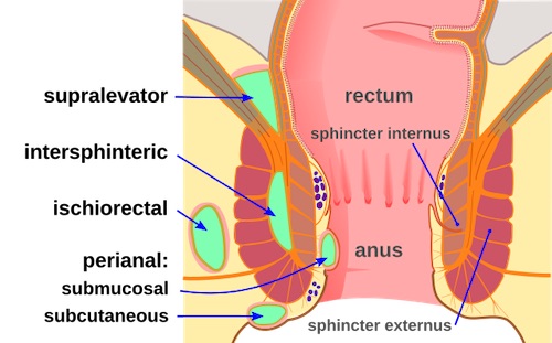
Anorectal Abscess Classification 1
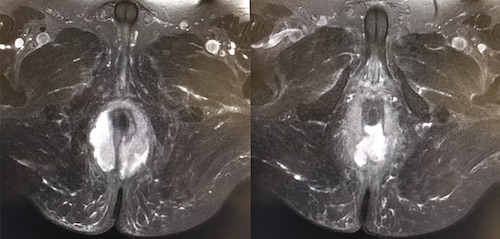
Horseshoe Abscess

Fistula-In-Ano

Basics

  • Most Common Cause: Cryptoglandular Disease
    • Infection Causes Abscess Leading to Fistula
  • External Opening Usually Obvious, Internal More Difficult to Identify
  • Goodsall’s Rule
    • Anterior Fistulas: Tract Radially to Nearest Crypt
      • Straight Path
    • Posterior Fistulas: Tract toward Posterior Midline
      • Curved Path

Classification

  • Route
    • Superficial/Subcutaneous: Does Not Involve the Sphincter Complex
    • Intersphincteric: Remains in the Intersphincteric Space
    • Transsphincteric: Passes Directly Through the EAS
    • Suprasphincteric: Extends Proximally in the Intersphincteric Space & Then Extends Through the Levators into the Ischiorectal Fossa
    • Extrasphincteric: Tract Extends from the Ischiorectal Fossa Through the Levators into the Rectal Wall
  • Complexity
    • Simple: No Complex Features
      • Superficial/Subcutaneous
      • Intersphincteric
      • Low Transsphincteric (< 30% of Sphincter Length)
      • Single Fistula
    • Complex:
      • High Transsphincteric Fistula (> 30% of Sphincter Length)
      • Suprasphincteric
      • Extrasphincteric
      • High Blind Tracts
      • Multiple Fistulas

Presentation

  • Drainage from External Opening – Mucoid, Bloody, Purulent or Feculent
  • Recurrent Abscess
  • Intermittent Anal Pain
  • Anal Pruritis
  • Anal Bleeding

Diagnosis

  • Primarily Based on History & Physical Exam
  • Simple Fistulas Require No Further Imaging
  • Complex Fistulas May Require Imaging to Guide Treatment
    • MRI or EUS are Preferred
    • Can Determine Presence/Course of Fistulous Tract, Localize Internal Opening, & Delineate Any Extensions
    • Imaging-Guided Surgery May Decrease Recurrence Rates for Complex Fistula

Treatment

  • Initial Management:
    • Simple: Fistulotomy
    • Complex: Seton
      • Consider Preoperative Transanal US or MRI to Define the Tract
      • Staged Procedure Once Tract is Matured (Simple, Narrow and Without Abscess)
    • Liberal Use of Setons in Setting of Crohn’s Disease
  • Other Options (Generally Used After Setons Fail):
    • LIFT Procedure (Ligation of Internal Fistula Tract)
    • Rectal Advancement Flap
    • Fibrin Glue – Low Success, Generally Abandoned
    • Collagen Plug – Low Success, Generally Abandoned
  • *See Anorectal: Abscess & Fistula Procedures

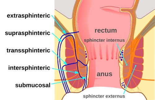
Anorectal Fistula Classification 1

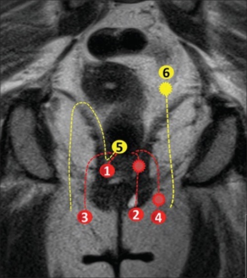
Anorectal Fistula Class by MRI: (1/2) Intersphincteric, (3/4) Transsphincteric, (5) Suprasphincteric, (6) Extrasphincteric 2
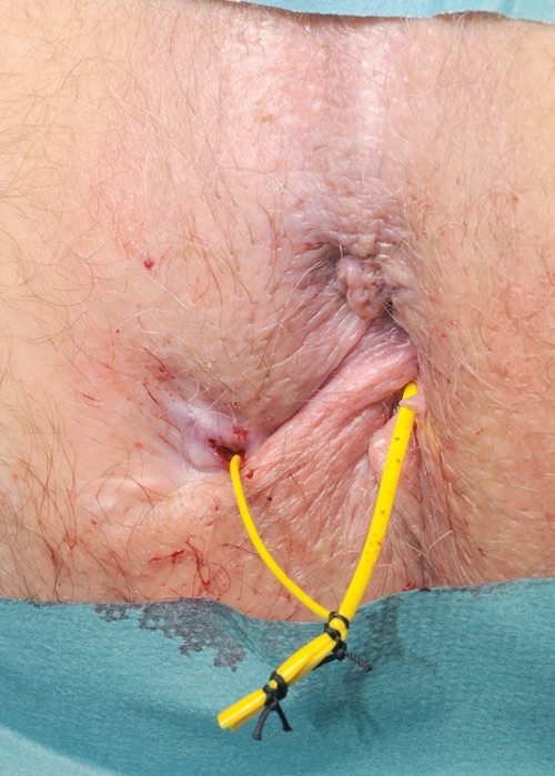
Seton 3

References

  1. Mcort NGHH. Wikimedia Commons. (License: CC BY-SA-4.0)
  2. Ram R, Sarver D, Pandey T, Guidry CL, Jambhekar KR. Magnetic resonance enterography: A stepwise interpretation approach and role of imaging in management of adult Crohn’s disease. Indian J Radiol Imaging. 2016 Apr-Jun;26(2):173-84. (License: CC BY-NC-SA-3.0)
  3. Tanner NC, Maw A. A novel technique for negotiation of a complex fistula-in-ano using a flexible ureteral catheter. Ann R Coll Surg Engl. 2014 Jan;96(1):80. (License: CC BY-3.0)