Prophylaxis
Treatment – First Episode
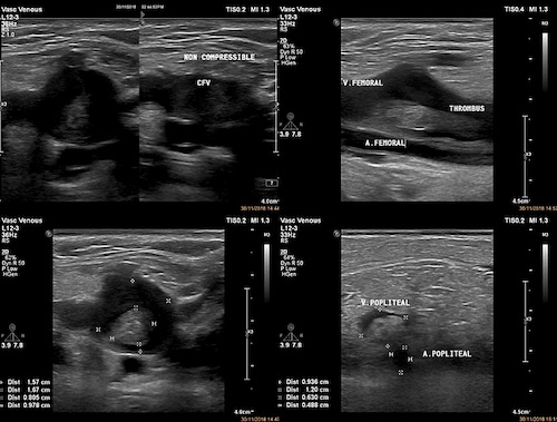
- Proximal DVT
- Provoked: 3-6 Months Anticoagulation
- Unprovoked: Long-Term (> 12 Months) Anticoagulation
- Consider Lifelong Anticoagulation if Hypercoagulable Disorder Present
- Distal DVT
- Symptomatic: Anticoagulation
- Asymptomatic: Serial US x2 Weeks
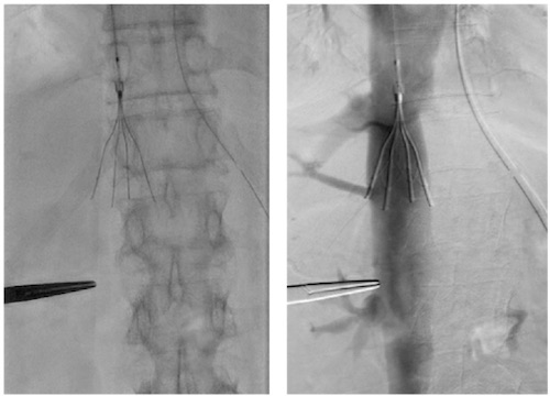
- Phlegmasia Cerulea Dolens
- Non-Threatened Extremity: Catheter-Directed Thrombolytics
- Threatened Extremity: Thrombectomy
- Choice of Agent:
- General Options:
- Unfractionated Heparin
- Low Molecular Weight Heparin (Lovenox)
- Fondaparinux
- Rivaroxaban
- Apixaban
- Warfarin/Coumadin – Cannot Be Sole Initial Treatment
- Malignancy: Low Molecular Weight Heparin (Lovenox)
- Pregnancy: Heparin or Low Molecular Weight Heparin (Lovenox)
- *May-Thurner Syndrome Managed with Venography, Thrombolysis/Thrombectomy & Left Iliac Stent
Treatment – Subsequent Episodes
- Second Episode: Long-Term (> 12 Months) Anticoagulation
- Third Episode: Life-Long Anticoagulation