Skin & Soft Tissue: Diabetic Foot Ulcer
Diabetic Foot Ulcer
Basics
- Up to 25% of Diabetics will Develop a Foot Ulcer During Their Lifetime
- Most Common Site: Second Metatarsophalangeal (MTP) Joint
- Also Common at Heel
- Over 50% Become Infected
- Can Progress to Osteomyelitis
Pathophysiology
- Peripheral Neuropathy
- Somatic Neuropathy
- Causes Loss of Protective Sensation
- Delayed Detection of Injury
- Autonomic Neuropathy
- Decreased Sweating with Dry & Fragile Skin
- Somatic Neuropathy
- Charcot Neuropathic Osteoarthropathy (Charcot Foot)
- “Rocker-Bottom” Foot with Midfoot Collapse
- Caused by Diabetes and Neuropathy
- Uncontrolled Inflammation Causes Osteolysis with Progressive Fracture and Dislocation
- Increases Risk of Ulceration from Altered Patterns of Mechanical Stress & Pressure
- Peripheral Arterial Disease – Causes Both Macrovascular & Microvascular Disease
- Macrovascular Changes – Infrapopliteal Occlusive Disease (Tibial and Peroneal Arteries)
- Generally Spares Aorto-Iliac and Femoral Arteries
- Microvascular Changes – Dysfunction of Autoregulation
- Endothelial Damage
- Increased Flow
- Edema
- Arteriovenous Shunting
- Impaired Oxygen Diffusion
- Impaired Leukocyte Migration
- Macrovascular Changes – Infrapopliteal Occlusive Disease (Tibial and Peroneal Arteries)
Risk Factors
- Peripheral Neuropathy – Most Common
- Peripheral Arterial Disease
- Immunosuppression
- Foot Deformity
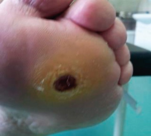
Diabetic Foot Ulcer 1
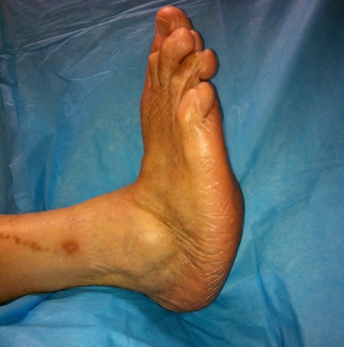
Charcot Foot 2
Evaluation
- Evaluate for Infection:
- Routine Culture Not Recommended
- Almost All are Colonized
- Consider Culture During Debridement
- Plain Radiograph is Often the First Step (Look for Bone Deformity or Necrotizing Infection)
- Use Sterile Probe to Interrogate for Any Abscess, Tendon or Bone
- Osteomyelitis:
- Do Not Biopsy Bone – Will Seed Bacteria into the Bone if Not Already Infected
- Can Confirm with MRI
- MRI Not Necessary if Bone is Exposed
- Routine Culture Not Recommended
- Evaluate for Ischemia:
- Ankle-Brachial Index (ABI)
- *See Vascular: Peripheral Arterial Disease (PAD)
- May Be Falsely Elevated Due to Arterial Calcification Seen in Diabetics
- Toe Pressure – Represents the Most Distal Microcirculation Pressure that Can Be Measured & Less Prone to Medial Calcinosis
- > 55 mmHg: Likely Able to Heal
- < 40 mmHg: Critical Ischemia
- Transcutaneous Oximetry (TcPO2)
- > 55 mmHg: Likely Able to Heal
- < 35 mmHg: Hypoxia, Indicates Need for Revascularization
- < 30 mmHg: Severe Ischemia
- < 10 mmHg: Critical Limb Ischemia
- Ankle-Brachial Index (ABI)
Treatment
- Optimize Glucose Control
- Offloading – Minimization of Weight Placed onto the Wound
- Consider Revascularization (Surgical vs Endovascular) if Indicated
- Debride Nonviable Tissue
- Acute Infection: Broad Spectrum Antibiotics
- Continue Antibiotics Until Infection is Cleared, Not Necessary Until Wound is Healed
- May Require Debridement or Amputation
- Strongly Consider Amputation for Osteomyelitis
Prevention
- Education
- Regular Foot Inspections
- Foot Hygiene
- Offloading Shoes
Diabetic Foot Ulcer – Classification
PEDIS Score
- PEDIS – Perfusion, Extent, Depth, Infection & Sensation
- Predicts 6-Month Risk of Amputation & Mortality
- Scoring:
- Perfusion
- No PAD: 0
- PAD without Critical Limb Ischemia: +1
- PAD with Critical Limb Ischemia: +2
- Extent
- Skin Intact: 0
- < 1 cm2: +1
- 1-3 cm2: +2
- > 3 cm2: +3
- Depth
- Skin Intact: 0
- Superficial: +1
- Fascia, Muscle or Tendon: +2
- Bone or Joint: +3
- Infection
- No Infection: 0
- Surface Infection: +1
- Abscess, Fasciitis or Septic Arthritis: +2
- Systemic Inflammatory Response Syndrome: +3
- Sensation
- Sensation Intact: 0
- Loss of Sensation: +1
- Perfusion
- Interpretation:
- Score < 7: Low Risk
- Score ≥ 7: High Risk
- 82% Specific for Nonhealing, Amputation or Death by 6-Months
Wagner Classification System
- Grade 0: No Ulceration
- Grade 1: Superficial Ulcer, Partial or Full Thickness
- Grade 2: Deep Ulcer Extending to Ligaments, Tendon, Joint Capsule or Deep Fascia
- Grade 3: Deep Ulcer with Abscess or Osteomyelitis
- Grade 4: Gangrene Localized to Forefoot or Heel
- Grade 5: Gangrene – Extensive
University of Texas Classification System
- Grade:
- 0: Pre-Ulcer or Post-Ulcer Epithelialization
- 1: Superficial Ulcer
- 2: Ulcer Penetrating to Tendon or Capsule
- 3: Penetrating to Bone or Joint
- Stage:
- A: No Infection or Ischemia
- B: Infection
- C: Ischemia
- D: Both Infection & Ischemia
WIfI Classification System
- WIfI – Wound, Ischemia, Foot Infection
- Scoring:
- Wound Grade
- 0: No Ulcer
- 1: Small Ulcer without Gangrene
- 2: Deep Ulcer or Gangrene Limited to Toes
- 3: Extensive Ulcer or Extensive Gangrene
- Ischemia Grade (Toe Pressure/TcPO2)
- 0: ≥ 60 mmHg
- 1: 40-59 mmHg
- 2: 30-39 mmHg
- 3: < 30 mmHg
- Foot Infection Grade
- 0: Uninfected
- 1: Mild (≤ 2 cm Cellulitis)
- 2: Moderate (> 2 cm Cellulitis or Purulence)
- 3: Severe (SIRS or Sepsis)
- Wound Grade
- Scores Used in a Staging Grid to Determine Risk of Amputation
References
- Zukic E, Gojak R, Novakovic A, Gazibera B. Predictive Role of Preventive Measures in Preventing the Progression of Diabetic Foot. Mater Sociomed. 2015 Aug;27(4):234-6. (License: CC BY-NC-4.0)
- Rogers LC, Frykberg RG, Armstrong DG, Boulton AJ, Edmonds M, Van GH, Hartemann A, Game F, Jeffcoate W, Jirkovska A, Jude E, Morbach S, Morrison WB, Pinzur M, Pitocco D, Sanders L, Wukich DK, Uccioli L. The Charcot foot in diabetes. Diabetes Care. 2011 Sep;34(9):2123-9. (License: CC BY-NC-ND-3.0)