Esophagus: Dysphagia & Stricture

Dysphagia

Causes

  • Oropharyngeal Dysphagia
    • UES Dysfunction
    • Neuromuscular Disease – Most Common Oropharyngeal Causes
      • Cerebrovascular Disease
      • Parkinson’s Disease
      • Neuropathy
      • Myasthenia Gravis
    • Oropharyngeal Carcinoma
    • Congenital Web
    • Zenker Diverticulum
  • Esophageal Dysphagia
  • Esophagogastric Dysphagia
    • Achalasia
    • Gastric Carcinoma
    • Stricture
  • Paraesophageal Dysphagia
    • Physical Obstruction (Thyromegaly, Left Atrial Enlargement, Postoperative Scarring, Lymphadenopathy)

Dakkak & Bennett Dysphagia Score

  • Grade 0: No Dysphagia
  • Grade 1: Dysphagia to Solids
  • Grade 2: Dysphagia to Semi-Solids
  • Grade 3: Dysphagia to Liquids
  • Grade 4: Aphagia

Diagnosis

  • Initial Test: Barium Swallow
    • Some Prefer to Preform Endoscopy First
  • Second Test: Upper Endoscopy
  • If No Structural Abnormality Seen: Esophageal Manometry

Diagnostics

Upper Endoscopy (Esophogastroduodenoscopy/EGD)

  • Best Initial Test for GERD (Visualize Esophagitis)
  • Not If Suspect Perforation

Esophagram

  • Barium Swallow
    • Pick Up Masses
    • Caustic to Tissue
    • Best Initial Test for: Dysphagia or Odynophagia
  • Gastrografin Swallow
    • Not Caustic
    • Best to Detect: Perforation

Manometry

  • Test Motility Disorders
  • Normal
    • UES Resting Pressure 60-80
    • LES Resting Pressure 6-26
    • LES Length > 2 cm
    • Abdominal Length > 1 cm

Strictures, Rings & Other Obstruction

Esophageal Stricture

  • Most Common Cause of Esophageal Dysphagia
  • Causes
    • Chronic GERD – Most Common
    • Radiation
    • Sclerotherapy
    • Caustic Injury
    • Pill-Induced Injury
    • Surgery
    • External Compression from Mediastinal Fibrosis
    • Eosinophilic Esophagitis
    • Malignancy
  • Types:
    • Simple: Short ≤ 2 cm, Straight & Luminal Diameter ≥ 12 mm
    • Complex: Long > 2 cm, Tortuous or Significant Narrowing (Diameter < 12 mm)
      • Often from Injury or Underlying Pathology
  • Primary Sx: Dysphagia
  • Tx: Esophageal Dilation
    • High Recurrence & Often Need Repeat Dilation
    • Dilation Techniques:
      • Bougie Dilators (Maloney)
        • “Rule of Three” – Never Pass More than Three Dilators of Sequential Size Once Moderate Resistance is Evident Per Session
      • Wire-Guided
      • Balloon/Pneumatic
    • Contraindications:
      • Absolute:
        • Acute Abdomen
        • Acute or Incomplete Healing of Perforation
      • Relative:
        • Bleeding Diathesis or Anticoagulant Use
        • Severe Pulmonary Disease or Recent MI
        • Large Thoracic Aneurysm
        • Pharyngeal or Cervical Deformity
        • Recent Laparotomy
    • If Continues to Fail After Multiple Dilations: Consider Esophagectomy

Esophageal Stricture 1

Schatzki Ring

  • Fibrous Narrowed Ring at the Squamous-Columnar Junction
  • Unknown Cause
  • Associated with GERD or Hiatal Hernia
  • Sx: Largely Asymptomatic but Can Cause Obstruction & Dysphagia
  • Tx:
    • ASx: Conservative Management
    • Sx: PPI & Pneumatic Dilation
      • Always Bx to Rule Out Malignancy

Esophageal Ring 2

Esophageal Web

  • Thin (< 2 mm) Eccentric Membrane
  • Most Common Site: Anterior Cervical Esophagus
  • Causes:
    • Plummer-Vinson Syndrome (Paterson-Brown-Kelley Syndrome)
      • Triad: Upper Esophageal Web, Dysphagia & Iron-Deficiency Anemia
    • Zenker’s Diverticulum
  • Tx: Pneumatic Dilation
    • Will Often Rupture During Endoscopy

Esophageal Web 3

Dysphagia Lusoria (Bayford-Autenrieth Dysphagia)

  • Latin for Dysphagia by a “Freak of Nature”/“Jest of Nature”
  • Definition: Dysphagia Caused External Compression from a Birth Defect of the Aortic Root Anatomy
  • Risk Factors: Down’s Syndrome & Congenital Heart Disease
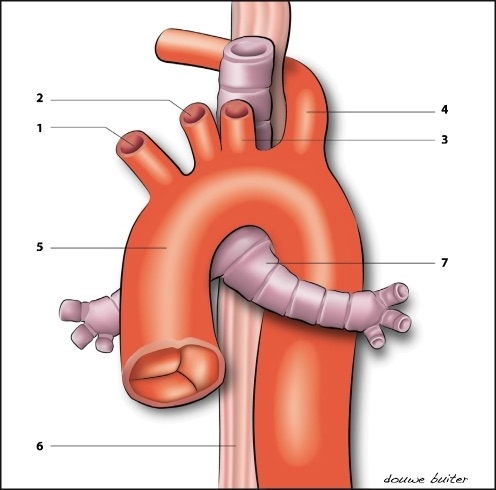
  • Abnormalities:
    • Aberrant Right Subclavian Artery off the Descending Aorta (After Left Subclavian)
      • Most Common Cause
      • The Primary Abnormality Described in the Literature
      • 60% Associated with an Aneurysm of the Proximal Portion (“Kommerell Diverticulum”)
    • Persistent Right Aortic Arch with Aberrant Left Subclavian Artery
    • Tortuous or Aneurysmal Thoracic Aorta
      • “Dysphagia Aortica”
    • Enlarged Left Atrium
  • Dx: CT Angiogram
    • Barium Esophagram Suggests but is Not Diagnostic
  • Tx:
    • Mild-Moderate Sx: Dietary & Lifestyle Modifications
      • Eat Slower with Small Bites & Chewing Well
    • Severe Sx: Vascular Reconstruction
      • If from Aberrant Right Subclavian Artery – Divide & Translocate the Distal Subclavian to the Aortic Arch or Right Common Carotid

Aberrant Right Subclavian 4

References

  1. Yamasaki Y, Ozawa S, Oguma J, Kazuno A, Ninomiya Y. Long peptic strictures of the esophagus due to reflux esophagitis: a case report. Surg Case Rep. 2016 Dec;2(1):64. (License: CC BY-4.0)
  2. Katsanos KH, Sigounas DE, Christodoulou DK, Tsianos EV. Upper esophageal ring due to gastric heterotopia. Ann Gastroenterol. 2012;25(2):163. (License: CC BY-NC-SA-3.0)
  3. Sánchez Prudencio S, Domingo Senra D, Martín Rodríguez D, Botella Mateu B, Esteban Jiménez-Zarza C, de la Morena López F, Jiménez Reyes J, Nevado Santos M, de Cuenca Morón B. Esophageal Cicatricial Pemphigoid as an Isolated Involvement Treated with Mycophenolate Mofetil. Case Rep Gastrointest Med. 2015;2015:620374. (License: CC BY-3.0)
  4. Roofthooft MT, van Meer H, Rietman WG, Ebels T, Berger RM. Down syndrome and aberrant right subclavian artery. Eur J Pediatr. 2008 Sep;167(9):1033-6. (License: CC BY-NC-2.0)