Cardiothoracic Surgery: Lung Cancer Presentation

Lung Cancer

Definitions

  • Solitary Pulmonary Nodule (SPN)/Coin Lesion: Single Lesion ≤ 3 cm
    • Causes:
      • Most Common Cause: Granuloma
      • Most Common Tumor: Hamartoma
      • Most Common Malignancy: Adenocarcinoma
    • 10% are Malignant Overall
      • Age < 40-50: < 5% are Malignant
      • Age > 50-60: > 50% are Malignant
  • Pulmonary Mass: Lesions > 3 cm
    • Higher Risk for Malignancy

General

  • Most Common Cause of Cancer-Related Death
  • Most Common Site of Mets: Brain
  • Strongest Prognostic Indicator: Nodal Metastases

Risk Factors

  • Smoking – Most Important Risk Factor (Accounts for 90% of All Lung Cancers)
    • Second Hand Smoke Increases Risk as Well
    • Cessation Can Decrease Risk by 20-90%
  • Lung Disease
    • COPD
    • Emphysema
    • Chronic Bronchitis
    • Pneumonia
    • Tuberculosis
    • Pulmonary Fibrosis
  • Radiation Therapy
  • Asbestos
  • Radon
  • Smoke from Cooking
  • Air Pollution

Types

  • Small Cell Lung Carcinoma (SCLC)
  • Non-Small Cell Lung Carcinoma (NSCLC)80% of Total Lung Cancers
    • Squamous Cell Carcinoma
    • Adenocarcinoma – Most Common Lung Cancer Overall & in Nonsmokers
    • Large Cell Carcinoma

Typical Location

  • Central Lesions: Mn
    • Small Cell Lung Carcinoma (SCLC)
    • Squamous Cell Carcinoma
  • Peripheral Lesions:
    • Adenocarcinoma
    • Large Cell Carcinoma

Prognosis

  • Small Cell Lung Carcinoma (SCLC)
    • Early-Stage Disease (< 5% – Rare): Stage I-II
    • Limited-Stage Disease (25%): Stage III
      • Median Survival: 15-20 Months
      • Five-Year Survival: 10-13%
    • Extensive-Stage Disease (75%): Stage IV
      • Median Survival: 8-13 Months
      • Five-Year Survival: 1-2%
  • Non-Small Cell Lung Carcinoma (NSCLC)
    • Five-Year Survival:
      • Stage I: 68-92%
      • Stage II: 53-65%
      • Stage III: 12-41%
      • Stage IV: 0-10%

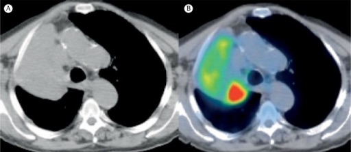
Non-Small Cell Lung Cancer on CT/PET 1

Presentation

Presentation

  • Persistent Cough – Most Common Presenting Symptom
  • Hemoptysis
  • Chest Pain
  • Dyspnea
  • Hoarseness
  • Pancoast Tumor – Tumor at the Apex of the Lung
    • Horner Syndrome (Invasion of Sympathetic Chain)
      • Sx: Ipsilateral Ptosis (Drooping Eyelid), Miosis (Pupil Constriction) & Anhidrosis (Minimal-No Sweat)
    • Brachial Plexus Involvement (Compression of C8 & T1 Nerve Roots)
      • Sx: Shoulder Pain & Symptoms Along the Ulnar Nerve Distribution
  • Superior Vena Cava (SVC) Syndrome
    • Compression of the SVC Due to an Extrinsic Source (Cancer Most Common)
    • Sx: Dyspnea & Swelling of the Head, Neck & Arms
    • CT Appearance: Nonopacification of SVC Inferior to Obstruction & Opacification of Chest Collaterals (Azygous/Intercostals)
  • Malignant Pleural Effusion

Paraneoplastic Syndromes

  • Small Cell Lung Carcinoma (SCLC)
    • Ectopic ACTH Secretion (Cushing Syndrome) – Most Common Paraneoplastic Syndrome Associated with SCLC
    • Syndrome of Inappropriate Antidiuretic Hormone Secretion (SIADH)
    • Lambert-Eaton Myasthenic Syndrome
    • Polymyositis & Dermatomyositis
  • Squamous Cell Carcinoma
    • Parathyroid Hormone (PTH)-Related Peptide Secretion Mn
      • *Also Seen in SCLC & Adenocarcinoma but Less Common
  • Adenocarcinoma
    • Hypercoagulability & Deep Venous Thrombosis (DVT)
    • Disseminated Intravascular Coagulopathy (DIC)
    • Superficial Thrombophlebitis (Trousseau Syndrome)
  • Large Cell Carcinoma
    • B-HCG Secretion – Gynecomastia

Pancoast Tumor 2

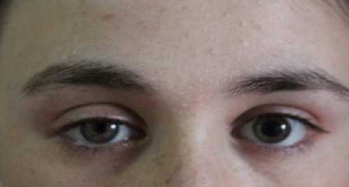
Horner Syndrome, Right-Side 3

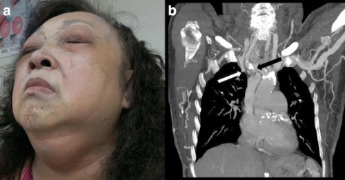
SVC Syndrome; (A) Facial Swelling, (B) CT Showing Occlusion (White Arrow) and Collaterals (Black Arrow) 4

Management

Mnemonics

Associations of Small Cell & Squamous Cell Cancer

  • S-S: Smokers
  • S-S: “Sentral”/Central

Paraneoplastic Syndrome of Squamous Cell Carcinoma

  • s-Ca-uamous: Calcium Increased from PTH-Related Peptide

References

  1. Hochhegger B, Alves GR, Irion KL, Fritscher CC, Fritscher LG, Concatto NH, Marchiori E. PET/CT imaging in lung cancer: indications and findings. J Bras Pneumol. 2015 May-Jun;41(3):264-74. (License: CC BY-NC-4.0)
  2. Upadhyaya V, Upadhyaya DN, Kumar A, Pandey AK, Gujral R, Singh AK. Magnetic resonance neurography of the brachial plexus. Indian J Plast Surg. 2015 May-Aug;48(2):129-37. (License: CC BY-NC-SA-3.0)
  3. Demiral M, Binay C, Simsek E, Ilhan H. Horner Syndrome Secondary to Thyroid Surgery. Case Rep Endocrinol. 2017;2017:1689039. (License: CC BY-4.0)
  4. Ho YJ, Yeh CH, Lai CC, Huang JC, Chuang LH. ExPRESS miniature glaucoma shunt for intractable secondary glaucoma in superior vena cava syndrome – a case report. BMC Ophthalmol. 2016 Jul 26;16:125. (License: CC BY-4.0)