Trauma: Neck Trauma

Zone Management

Zones of the Neck

  • Zone I: Thoracic Inlet to Cricoid Cartilage
  • Zone II: Cricoid Cartilage to Angle of Mandible
    • Most Vulnerable to Injury
    • Most Commonly Injured
  • Zone III: Angle of Mandible to Base of Skull

Signs of Injury

  • Hard Signs: Definitive Indications of Arterial/Structural Injury
    • Active Hemorrhage
    • Expanding or Pulsatile Hematoma
    • Subcutaneous Emphysema or Air Bubbling from Wound
  • Soft Signs: Possible Indications of Injury
    • Dysphagia
    • Voice Changes
    • Hemoptysis

Initial Management

  • Stable & No Hard Signs: CTA
    • Penetrating Specific Considerations:
      • If Platysma Not Violated: Observation
      • Zone I – Consider CXR First
      • Zone II – Some Consider Surgical Exploration First without CT (*Classic Teaching)
  • Unstable, Hard Signs or Symptomatic: Surgical Exploration
    • Incision is Made Along the Anterior Border of the Sternocleidomastoid Muscle (SCM) – Similar to a Carotid Endarterectomy
    • If Bilateral Exploration is Required – Can Make Bilateral Incisions that Connect Inferiorly in a “U”-Shape

Gaining Vascular Control at Surgery

  • Zone I – Proximal Control Requires Median Sternotomy
    • Left Subclavian Injury May Require Anterior Left Thoracotomy
  • Zone II – Directly Control
  • Zone III – Distal Control Requires Mandible Disarticulation vs Antegrade Embolectomy Catheter
    • Most Challenging

Zones of the Neck

Vascular Injury

Blunt Cerebrovascular Injury (BCVI)

  • Indications for CTA (2010 EAST Guidelines):
    • Level II Evidence:
      • Neurologic Injury Not Explained by a Diagnosed Injury
      • Epistaxis from a Suspected Arterial Source
    • Level III Evidence:
      • GCS ≤ 8
      • Petrous Bone Fracture
      • DAI
      • C-Spine Fracture – Especially C1-C3 & Through the Foramen Transversarium
      • C-Spine Fracture with Subluxation
      • LeFort II or III Facial Fractures
  • *Seatbelt Sign Alone is Not an Indication for CTA – Should be Reserved for Those with other Associated Clinical Findings
  • Biffl/Denver Scale
    • Grade I: Mild Intimal Irregularity < 25% Narrowing
    • Grade II: Dissection or Intramural Hematoma > 25% Narrowing
    • Grade III: Pseudoaneurysm
    • Grade IV: Occlusion or Total Thrombosis
    • Grade V: Transection
  • Treatment:
    • Grade I: ASA
    • Grade II-IV: Heparin
      • Intervention Indication (Endovascular Stent vs Surgery):
        • Grade II: Neurologic Sx
        • Grade III: Sx or > 1.0 cm
        • Grade IV: Sx or Evolving
    • Grade V: Surgical Repair
      • Short Segment: Primary Anastomosis
      • Large Segment: Saphenous Vein Graft
      • Unstable: Ligation

BCVI Grade II 1

BCVI Grade III 1

BCVI Grade IV 2

Right CCA Transection 3

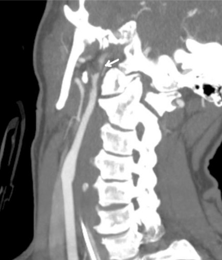
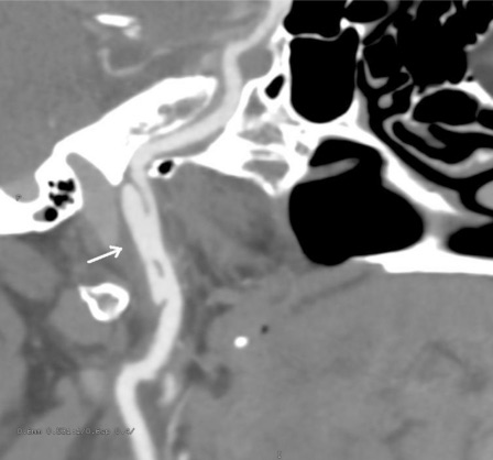
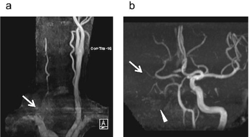
Internal Carotid Artery – Penetrating Injury

  • Tx: Surgery
  • Surgical Repair Options:
    • Primary Arteriorrhaphy
    • Patch Angioplasty
    • End-to-End Anastomosis
    • Vein or PTFE Graft
    • ECA Transposition to Injured ICA
  • Ligation Indications (High CVA Risk: 75-80%):
    • Unstable
    • Very Severe Neck Injury
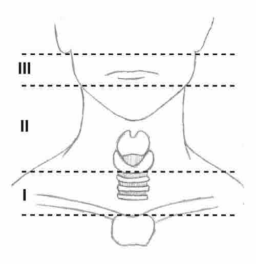
    • Zone III ICA Injury at Skull Base

External Carotid Artery

  • Safe to Ligate if Needed

Internal Jugular Vein

  • Transverse Venorrhaphy if Able
  • Major Hemorrhage/Unstable: Ligate

Subclavian/Axillary Artery

  • Tx: Endovascular vs Open Repair
    • Pseudoaneurysm: Endovascular Stent
    • Laceration > 50%/Transection: Open Repair

Other Injury

Esophagus

Larynx/Trachea

  • Dx: Laryngoscopy/Bronchoscopy
    • Can Be Evaluated in the OR if Unstable or There for Other Reasons
  • Small: Repair Transversely in 1-Layer with Absorbable Suture
    • 2-Layers Risk Stenosis
    • Include Tracheal Rings in Repair
  • Large: Primary Anastomosis (Up to 5-6 Tracheal Rings in Length)
    • Strongly Consider Tracheostomy – Place in Standard Position or Possibly Through the Injured Site

Recurrent Laryngeal Nerve

  • Tx: Repair or Re-Implant in Cricoarytenoid Muscle

Thyroid

  • Tx: Control Bleed & Drain (No Resection)

Larynx Laceration

References

  1. Karamchandani R, Rajajee V, Pandey A. The role of neuroimaging in the latent period of blunt traumatic cerebrovascular injury. Open Neuroimag J. 2011;5:225-31. (License: CC BY-NC-3.0)
  2. Kobayashi K, Imagama S, Okura T, Yoshihara H, Ito Z, Ando K, Ukai J, Shinjo R, Muramoto A, Matsumoto T, Nakashima H, Ishiguro N. Fatal case of cervical blunt vascular injury with cervical vertebral fracture: a case report. Nagoya J Med Sci. 2015 Aug;77(3):507-14.(License: CC BY-NC-ND-4.0)
  3. Babu A, Garg H, Sagar S, Gupta A, Kumar S. Penetrating neck injury: Collaterals for another life after ligation of common carotid artery and subclavian artery. Chin J Traumatol. 2017 Feb;20(1):56-58. (License: CC BY-NC-ND-4.0)