Skin & Soft Tissue: Plastics & Tissue Transfer

Definitions

Tissue Transfer Definitions

  • Flap: Tissue Transfer with an Intact Blood Supply
  • Graft: Tissue Transfer without an Intact Blood Supply

Complex Defect Reconstructive Ladder (Simple to Complex)

  • Secondary Intention
  • Primary Closure
  • Skin Graft
  • Local Flap
  • Regional Flap
  • Free Flap

Flaps

Blood Supply

  • Pedicled Flap: Pedicle of Donor Site with Vascular Supply Remains Intact
  • Free Flap: Tissue Completely Detached from Donor Site
    • Blood Supply Reconstituted at Recipient Site by Microsurgery
    • Microvascular Free Flap Survival: 95%

Proximity

  • Local Flap: Uses Tissue That Abuts the Defect
    • Types
      • Advancement Flap: Tissue Stretched in a Straight Line
        • Ex: V-Y Flap
      • Rotational Flap: Tissue Stretched in an Arc
      • Transposition Flap: Tissue Rotated, Creating a New Defect
        • Ex: Rhomboid Flap or Z-Plasty
  • Regional Flap: Uses Tissue Near the Defect that Does Not Actually Abut the Defect
    • Still Connected to Donor Site by Pedicle
    • Interpolation Flap: A 2-Stage Regional Flap
      • First the Flap is Transferred Keeping a Pedicle Connected to the Donor Site Over/Under a Skin Bridge
      • Later the Pedicle is Removed After Neovascularization
  • Distant Flap: Uses Tissue Far from the Defect
    • Can Be Pedicled or Free Flap

Advancement Flap 1

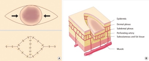
V-Y Advancement Flap 2
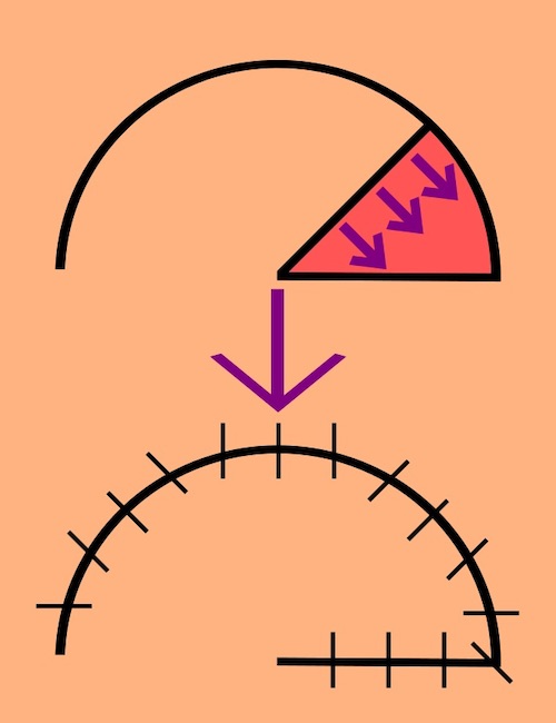
Rotational Flap 1

Transposition Flap 1
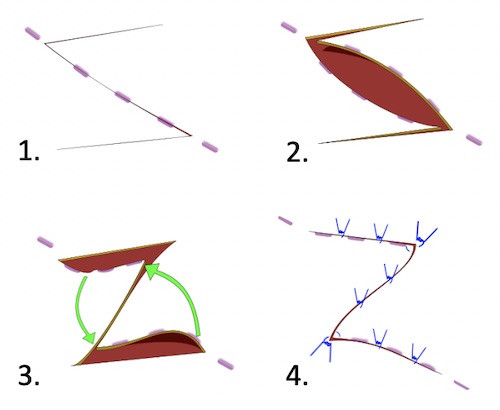
Z-Plasty 3

Common Flaps

  • Breast Reconstructions:
    • Transverse Rectus Abdominis Myocutaneous (TRAM) Flap
    • Deep Inferior Epigastric Perforator (DIEP)/Fasciocutaneous Flap
    • Superficial Inferior Epigastric Artery (SIEA) Flap
    • Latissimus Dorsi Myocutaneous Flap (LDMF)
    • Thoracodorsal Artery Perforator Flap (TDAP)
    • *See Breast: Breast Reconstruction
  • Other Common Flaps:
    • Forehead Flap
    • Omental Flap
    • Anterolateral Thigh Flap
    • Radial Forearm Flap
    • Medial Gastrocnemius Flap

Grafts

Types of Grafts

  • Autograft
    • Definition: Graft from the Same Patient Themselves
    • Best Outcomes
  • Allograft/Homograft
    • Definition: Graft from Another Human or Cadaver
      • Decellularized & Rendered Nonimmunogenic by Washing Process
    • Will Vascularize
    • Temporize (4 Weeks) – Will Eventually Reject & Must be Replaced
  • Xenograft
    • Definition: Acellular Collagen Extracts from Nonhuman Sources (Porcine or Bovine)
    • No Vascularization
    • Temporize (2 Weeks)
  • Dermal Substitute
    • Uses Synthetic Materials (Vicryl, PTFE, Prolene, etc.)
    • Worst Outcomes

Autograft Thickness

  • Split-Thickness Skin Graft (STSG)
    • Layers: Epidermis & Superficial Dermis
    • Most Likely to Survive (Thin with Better Imbibition)
    • Donor Site Heals by Edges & Hair Follicles After 1-2 Weeks
    • Harvest with a Dermatome
      • If Too Deep: Stop, Suture Repair & Start at Another Site
    • Meshing:
      • Increases Tissue Coverage & Decreases Amount Harvested
      • Allows Drainage of Fluid/Blood (Lower Seroma/Hematoma Risk)
      • Heals by Secondary Intention Between Gaps
        • Increased Scarring & Longer Healing Time
  • Full-Thickness Skin Graft (FTSG)
    • Layers: Epidermis & All of Dermis (No Subcutaneous)
      • Remove Fat to Avoid Fat Necrosis & Facilitate Revascularization
    • Less Contraction
    • Generally Only Used for Small Areas for Cosmetics/Function (Such as Face or Hand)
    • Potential Donor Sites: Post-Auricular, Supraclavicular, Lateral Neck, Inner Arms, Forearms & Inguinal Crease
      • Choose Site with Best Cosmetic Match
    • Harvest with a Knife & Close Donor Site Primarily (Will Not Heal)

Graft Survival

  • Initial: Imbibition
    • Osmotic Diffusion
  • Day 3-4: Inosculation
    • Direct Connection to Wound Bed Vessels
  • Day 5: Neovascularization
    • Formation of New Blood Vessels with Angiogenesis

Contraction

  • Primary Contraction
    • Occurs Immediately at the Time of Harvest
    • Caused by Elastic Recoil of Dermis
      • Directly Proportional to the Amount of Dermis
    • Higher in FTSG
  • Secondary Contraction
    • Delayed Contraction
    • Caused by Healing Skin & Host Bed Contraction
      • Inversely Proportional to the Amount of Dermis
    • Higher in STSG

Causes of Graft Failure

  • Hematoma/Seroma of Wound Bed – Most Common Cause
    • Prevents Nutrient Delivery & Neovascularization
  • Infection
  • Poor Wound Bed/Vascularity
  • Sheer Forces

Donor Site Management

  • Must Achieve Adequate Hemostasis – High Risk for Significant Blood Loss
  • Moist Environments are Generally Preferred
    • Options: Xeroform, Petroleum-Impregnated Gauze or Alginate
    • Moist Environments Improve Wound Healing & Decrease Pain
    • Dry Gauze Causes Pain When Changing
    • Leaving Open to Air Increases Risk of Infection
  • Scarring is Expected
  • Donor Site is Generally More Painful than the Recipient Site

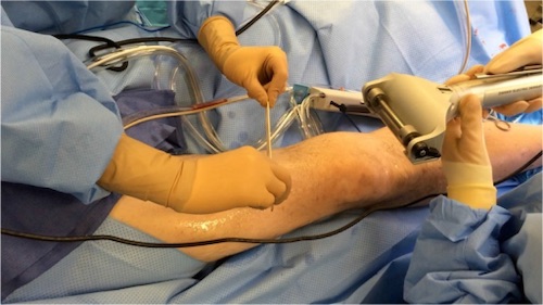
Skin Graft Dermatome 4

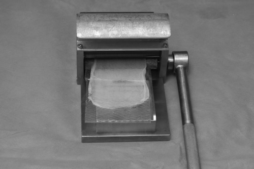
Dermacarrier & Mesher 5
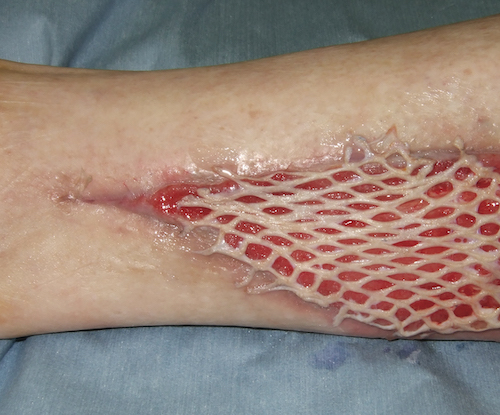
Meshed STSG 6

References

  1. Taylornate. Wikimedia Commons. (License: CC BY-SA-3.0)
  2. Kwon KH, Lee DG, Koo SH, Jo MS, Shin H, Seul JH. Usefulness of v-y advancement flap for defects after skin tumor excision. Arch Plast Surg. 2012 Nov;39(6):619-25. (License: CC BY-NC-3.0)
  3. Mason E. Wikimedia Commons. (License: CC BY-SA-4.0)
  4. Miller JD, Rankin TM, Hua NT, Ontiveros T, Giovinco NA, Mills JL, Armstrong DG. Reduction of pain via platelet-rich plasma in split-thickness skin graft donor sites: a series of matched pairs. Diabet Foot Ankle. 2015 Jan 22;6:24972. (License: CC BY-NC-ND-3.0)
  5. Wokes JE, Ali-Khan A. Microfenestrated split thickness skin grafts: an underused technique? Ann R Coll Surg Engl. 2012 Sep;94(6):447. (License: CC BY-3.0)
  6. Sakamoto, Y., & Kishi, K. (2013). The Fixation and Dressing for Meshed and Sheet Skin Graft. In (Ed.), Skin Grafts. IntechOpen. (License: CC BY-3.0)