Trauma: Pulmonary Trauma

Pulmonary Injury

AAST Lung Injury Scale

Pulmonary Contusion

  • Edema & Inflammation Causing Impaired Gas Exchange
  • Most Common in High-Energy Blunt Trauma
  • Presents 24-48 Hours Post-Trauma
    • CXR Changes Evolve Slowly & Lag Behind Worsening Respiratory Status
  • Dx: CXR/CT
  • Tx: Diuretics & Fluid Restriction

Pulmonary Parenchymal Injury

  • Large Laceration
    • Tx: Wedge Resection
  • Deep Penetrating Wound with Air Leak/Bleeding
    • Tx: Tractotomy & Deep Vessel Ligation
      • Linear Stapler Through Tract to Access Bleeding Vessel
  • Devascularized/Destroyed Lobe
    • Tx: Lobectomy
  • Last Resort: Pneumonectomy
    • Complications: Severe Respiratory Failure and Right-Sided Heart Failure

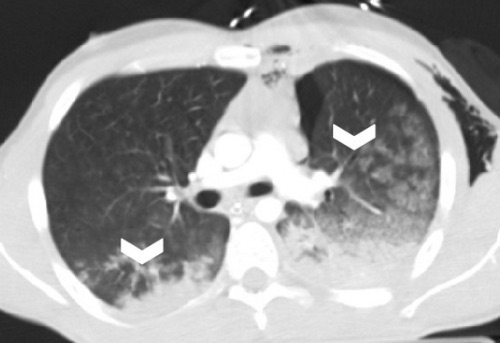
Pulmonary Contusion 1

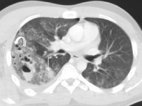
Pulmonary Laceration; Curved Arrow – Pneumatocele, Straight Arrow – Hematocele 1

Pneumothorax (PTX)

Nontraumatic

CXR Findings

  • Sensitivity:
    • Upright: 92%
    • Supine: 50%
    • *20% of “Occult” PTX in Trauma was Actually “Missed”
  • General Radiographic Findings:
    • Pleural Line Separating Lung from Chest Wall
    • Loss of Normal Lung Markings to the Peripheral Chest Wall
  • Radiographic Signs in a Supine CXR:
    • Deep Sulcus Sign: Abnormally Deepened Ipsilateral Costophrenic Angle
      • Seen on Supine CXR Because Air Collects Anteriorly/Basally
      • The Most Commonly “Missed” Sign
      • COPD Can Cause a False Deep Sulcus Sign Due to Hyperinflation
    • Relative Lucency of Ipsilateral Hemithorax
    • Increased Sharpness of Cardiac Border, Mediastinal Margin or Diaphragm
    • Depression of the Ipsilateral Hemidiaphragm
    • Double Diaphragm Sign: Visualization of the Anterior Costophrenic Sulcus (Due to Air) and the Diaphragmatic Dome (Due to Lung Border)
    • Pericardial Fat Tag Sign: Nodular Contour of the Cardiac Border
      • Due to Change in Shape of the Pericardial Fat that is No Longer Compressed by the Lung

Simple Pneumothorax

  • After Penetrating Injury 80% Have Concomitant HTX
  • Dx: CXR/CT
  • PEEP from Mechanical Ventilation Can Cause Persistent Air Leaks
  • Tx:
    • ASx & Small (< 3 cm): Repeat CXR in 6 Hours
      • Supplemental Oxygen May Enhance Reabsorption – Reduces Partial Pressure of Nitrogen for a Diffusion Gradient
    • Sx or > 3 cm: Chest Tube

Tension Pneumothorax

  • Sx: JVD, Hypotension & Tracheal Deviation
  • Underlying Pathophysiology: Decreased Venous Return & Circulatory Collapse
  • Dx: Clinical
  • Tx: Needle Decompression Followed by Chest Tube
    • Large Bore Needle (14 Gauge) Through Midaxillary Line at 4th/5th Intercostal Space
    • *Historically Placed at the 2nd/3rd Intercostal Space – Higher Risk of Failing to Actually Penetrate the Pleural Cavity

Open Pneumothorax (Sucking Chest Wound)

  • Opens on Inspiration, Closes on Expiration
  • Can Cause Tension PTX
  • Tx: Chest Tube (Distant Site) & Occlusive Dressing, Taped on 3 Sides
    • Allows Air to Escape but Not Enter

PTX

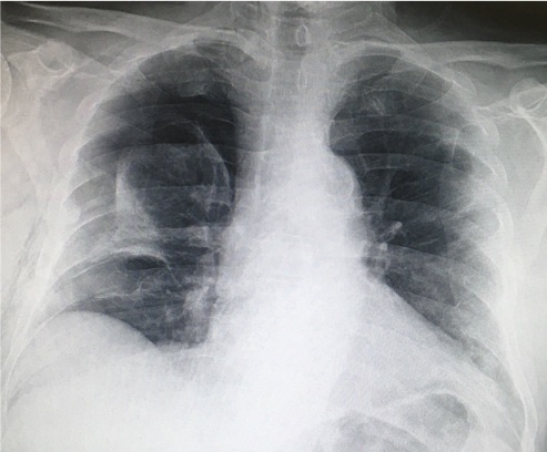
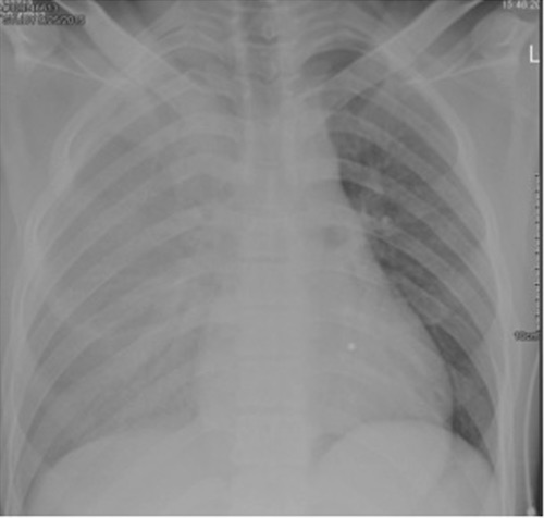
Deep Sulcus Sign (Right) on Supine CXR 2

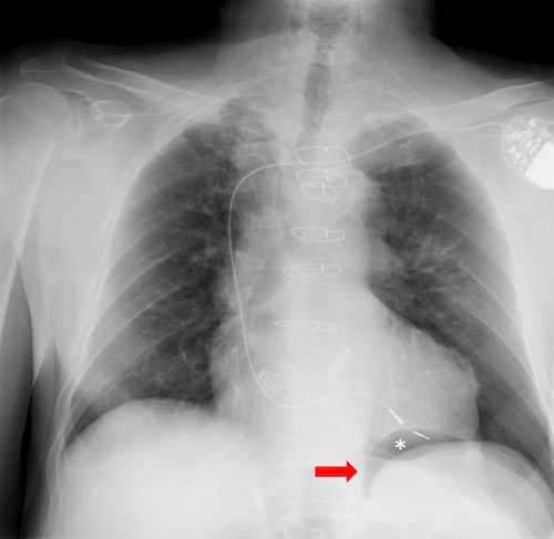
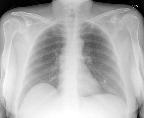
Double Diaphragm Sign (Astrisk) on Supine CXR 3

Hemothorax (HTX)

Treatment

  • Initial Tx: Chest Tube
  • Thoracotomy Indications:
    • Initial Loss: > 1,500 cc
    • Continual Loss: > 200 cc/hr for 4 Hours
      • Some Say 250 cc/hr for 3 Hours
    • If Concern for Major Vessel Injury: Preform Appropriate Arterial Exposure
  • Second Chest Tube May Be Required if Not Draining (Not After 1-2 Days)

Retained HTX

  • Chest Tube Fails to Completely Evacuate HTX After 2-3 Days
  • Occurs in 5% of Cases
  • Risk Factors: Prolonged Ventilation, PNA or Break in Pleura (Chest Tube)
  • High Risk of Fibrothorax and Empyema
  • Dx: CT (Need Before OR)
  • Tx: VATS
    • Not Second Chest Tube
    • Timing: Preform Within 7 Days (Before Loculations Develop)

HTX 4

Retained HTX with Hematocrit Sign 1

Tracheobronchial Injury

Basics

  • If SpO2 Drops After Chest Tube – Clamp Chest Tube
  • Most Common on Right & with Blunt Trauma
    • 90% within 1 cm of Carina

Diagnosis

  • Presentation: Large Continuous Air Leak, Hemoptysis, Pneumomediastinum & PTX
  • Dx: Bronchoscopy

Airway Management

  • Maintaining Airway: Consider Conservative
  • Suspected Laryngotracheal Injury: Tracheostomy
    • Endotracheal Intubation Can Worsen Damage
  • Distal Injury: Mainstem Single-Lumen Endotracheal Tube to the Unaffected Side

Surgical Repair

  • Indications:
    • Respiratory Compromise
    • Unable to Insufflate Lung
    • Injury > 1/3 Diameter of Trachea
    • Persistent > 2 Weeks
  • Approach:
    • Extrathoracic Injury: Transverse Cervical Incision
    • Most Thoracic Injuries: Right Posterolateral Thoracotomy
    • Distal Left Mainstem Injury (> 3 cm from Carina): Left Posterolateral Thoracotomy
  • Surgery: Primary Repair or End-to-End Anastomosis
    • If Under Tension: Can Flex Neck & Suture Chin to Chest to Maintain Postoperatively
    • Consider Reinforcement with Vascularized Pedicle or Intercostal or Strap Muscles

Left Main Bronchus Transection 5

Bronchial Transection 6

References

  1. Palas J, Matos AP, Mascarenhas V, Herédia V, Ramalho M. Multidetector computer tomography: evaluation of blunt chest trauma in adults. Radiol Res Pract. 2014;2014:864369. (License: CC BY-4.0)
  2. Kohli MD, Rosenman M. Indiana University.2013. (License: CC BY-NC-ND-4.0)
  3. Moreno SA, et al. Thoracic Trauma: A Comprehensive Review of Radiological Findings. ECR 2015. Poster C-2031. DOI: 10.1594/ecr2015/C-2031. (License: Open Liscence – Not Specified)
  4. Mishra B, Joshi MK, Kumar S, Kumar A, Gupta A, Rattan A, Sagar S, Singhal M, Misra MC. Innocuous cardiac gunshot that proved fatal: A bitter lesson learned. Chin J Traumatol. 2017 Apr;20(2):122-124. (License: CC BY-NC-ND-4.0)
  5. Groenendijk MR, Hartemink KJ, Dickhoff C, Geeraedts LM Jr, Terra M, Thoral P, Hashemi SM. Pneumomediastinum and (bilateral) pneumothorax after high energy trauma: Indications for emergency bronchoscopy. Respir Med Case Rep. 2014 Jul 11;13:9-11.(License: CC BY-NC-ND-3.0)
  6. Oikonomou A, Prassopoulos P. CT imaging of blunt chest trauma. Insights Imaging. 2011 Jun;2(3):281-295. (License: CC BY-4.0)