Breast: Proliferative Lesions

Proliferative Lesions

Lesions without Atypia

  • Fibroadenoma
  • Intraductal Papilloma
  • Usual Ductal Hyperplasia
  • Benign Sclerosing Lesions:
    • Radial Scar (RS): < 1 cm
    • Complex Sclerosing Lesion (CSL): > 1 cm
  • Sclerosing Adenosis – Lobular Lesion with Increased Fibrous Tissue & Interspersed Glandular Tissue

Lesions with Atypia

  • Atypical Ductal Hyperplasia (ADH)
  • Atypical Lobular Hyperplasia (ALH)

Risk for Upgrade to Malignancy After Excision

  • Low:
    • Fibroadenoma
    • Usual Ductal Hyperplasia
    • Sclerosing Adenosis
  • Intraductal Papilloma: 5-15%
  • Benign Sclerosing Lesions (RS/CSL): 8-20%
  • Atypical Ductal Hyperplasia (ADH): 5-10%
  • Atypical Lobular Hyperplasia (ALH): 2.5%

Diagnosis

  • Initial Evaluation: Imaging (Mammogram/US)
  • Diagnosis: Core Needle Biopsy (CNB)
  • Specifics for Benign Sclerosing Lesions:
    • Imaging May Demonstrate Stellate Lesions with Central Translucency/Sclerosis & Entrapped Peripheral Ducts/Lobules
    • Pathology: Fibroelastic Core with Distorted/Stellate Ducts
  • *See Breast: Breast Mass Evaluation

Treatment

  • Most Lesions without Atypia Require No Further Intervention
  • Indications for Surgical Excision:
    • Atypia (ADH/ALH)
      • May Consider Avoiding Excision for ALH if Imaging & Biopsy are Concordant
    • Benign Sclerosing Lesions (Radial Scar/Complex Sclerosing Lesion)
    • Intraductal Papilloma
  • Margins: No Margin Required
    • Goal: All Suspicious/Calcified Areas on Mammogram

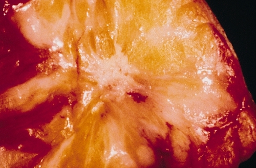
Radial Scar; Gross Pathology 1

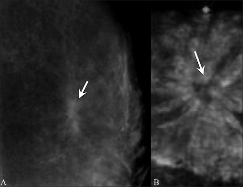
Radial Scar; (A) Mammogram, (B) 3D Multiplanar US 2

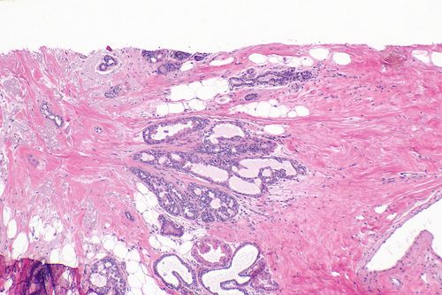
Radial Scar; Histology 3

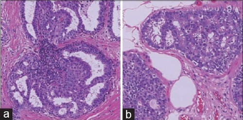
Intraductal Lesions: (A) Usual Ductal Hyperplasia, (B) Atypical Ductal Hyperplasia 4

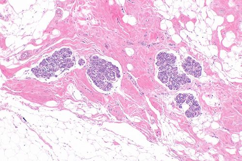
Atypical Lobular Hyperplasia 3

References

  1. The Armed Forces Institute of Pathology. Wikimedia Commons. (License: Public Domain)
  2. Shah G, Jankharia B. Pictorial essay: Breast USG. Indian J Radiol Imaging. 2010 May;20(2):98-104. (License: CC BY-2.0)
  3. Nephron. Wikimedia Commons. (License: CC BY-SA-4.0)
  4. Yamada M, Saito A, Yamamoto Y, Cosatto E, Kurata A, Nagao T, Tateishi A, Kuroda M. Quantitative nucleic features are effective for discrimination of intraductal proliferative lesions of the breast. J Pathol Inform. 2016 Jan 29;7:1. (License: CC BY-NC-SA-3.0)