Vascular: Acute Limb Ischemia (ALI)
Etiology
Causes of Acute Limb Ischemia
- Trauma
- Iatrogenic
- Arterial Thrombosis
- Arterial Embolism
Thrombosis
- Definition: Local Blood Clot that Develops Within the Circulatory System
- In Extremities May Be Bilateral
- Typically More Chronic with History of Claudication & Collaterals
- Can Occur in Grafts
- Peripheral Causes:
- Atherosclerosis
- Hypercoagulability
- Arterial Dissection
Embolism
- Definition: Distant Lodging of Material Causing Obstruction of a Vessel
- Secondary Thrombus forms Around Emboli & Exacerbates
- Typically Acute with No History of Claudication & No Collaterals
- Emboli:
- Thromboembolism – Blood Clot
- Most Common Source: Atrial Fibrillation Causing Thrombi in the Left Atrial Appendage
- Atheroma Embolism – Cholesterol Plaques that Embolize & Lodge Distally
- Can Be Spontaneous or Caused by Intraarterial Manipulation
- Most Common Site: Renal Arteries
- Embolic Blue Toe Syndrome
- Ischemic Toes from Atheromatous Embolism
- Most Common Source: Aortoiliac Disease
- Air Embolism – *See Vascular: Other Venous Emboli
- Fat Embolism – *See Vascular: Other Venous Emboli
- CO2 Embolism – *See Surgical Principles: Minimally Invasive Surgery (MIS)
- Endocarditis
- Atrial Myxoma
- Bullet Embolism – *See Trauma: Thoracic Vascular Trauma
- Thromboembolism – Blood Clot
- Most Common Sites: At Bifurcations
- Overall & Lower Extremity: Common Femoral Artery (At Bifurcation)
- Upper Extremity: Brachial Artery (Distal)

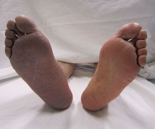
Acute Limb Ischemia, Cyanotic Right Foot 1
Presentation & Diagnosis
Presentation
- Absent Peripheral Pulses
- First Affects Sensory Nerves (Paresthesia & Loss of Sensation)
- Next Affects Motor Nerves (Paresis & Paralysis)
- Next Affects Skin
- Initial Pallor
- Then Dusky Blue from Capillary Venodilation
- Pressure Leaves Blush/White as Vessels are Empty
- Finally Pressure Produces No Blush Due to Capillary Extravasation
- Finally Affects Muscles (Muscle Tenderness) – Late Sign
- 6 P’s – Similar to Compartment Syndrome
- Pain
- Paresthesia (Pins & Needles Sensation)
- Paresis (Weakness) or Paralysis (Unable to Move)
- Pallor (Pale Color)
- Poikilothermic (Cold)
- Pulseless
Complications
- Ischemia-Reperfusion Injury
- Compartment Syndrome
- Permanent Nerve Damage
- Limb Loss
- Death
Diagnosis
- Clinical Diagnosis is Sufficient if Classic History & Physical Exam
- Imaging Options:
- CT Angiogram – Often the Test of Choice
- Duplex Ultrasound
- Transfemoral Arteriography

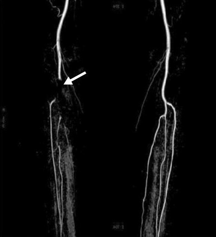
Acute Occlusion of the Popliteal Artery 2
Rutherford Acute Limb Ischemia Class
| Class | Sensory Function | Motor Function | Arterial Doppler Signal | Venous Doppler Signal |
| I – Viable | Normal | Normal | Audible | Audible |
| IIa – Marginally Threatened | Loss to Toes | Normal | Inaudible | Audible |
| IIb – Immediately Threatened | Loss Beyond Toes & Rest Pain | Weakness | Inaudible | Audible |
| III – Irreversible (Unsalvageable) | Complete Loss | Complete Loss | Inaudible | Inaudible |
Treatment
Initial Treatment
- Anticoagulation (Heparin) for All Patients
- Bolus & Drip
- Stabilizes Clot to Prevent Secondary Thrombosis but Has No Direct Thrombolytic Effect
- Other Measures: Supplemental Oxygen, IV Fluids & Analgesia
Selection & Timing
- Class I: Medical Management
- Class IIa: Urgent Intervention
- Class IIb: Emergent Intervention for Limb Salvage
- Class III: Do Not Revascularize
Definitive Treatment
- Class I (Viable)
- Medical Management with Anticoagulation Alone
- Class IIa (Marginally Threatened)
- Consider Open Surgery vs Endovascular Interventions
- Endovascular Indications:
- Short Duration (< 2 Weeks)
- Bypass Graft Occlusions
- Poor Surgical Options
- Occluded Runoff Vessels
- Surgery Indications:
- Long Duration (> 2 Weeks)
- Patient is Fit with a Good Surgical Option
- Class IIb (Immediately Threatened)
- Typically Open Thromboembolectomy
- May Consider Thrombolysis with High-Dose Bolus Infusion in Experienced Units if Quickly Available
- Class III (Irreversible/Unsalvageable)
- Amputation
- Do Not Revascularize – Futile with Systemic Complications
Endovascular Interventions
- Percutaneous Thrombolytics (tPA or Urokinase)
- Administration:
- Catheter-Directed to Clot to Minimize Systemic Effects
- Check Fibrinogen Every 4-6 Hours – Hold if Fibrinogen < 100 mg/dL
- Must Remain NPO & Bedrest to Observe
- Thrombolytic Contraindications:
- Absolute:
- Active Bleeding
- GI Bleed ≤ 10 Days
- CVA ≤ 6 Months
- Head Injury ≤ 3 Months
- Intracranial/Spinal Surgery ≤ 3 Months
- Relative:
- CPR, Major Surgery or Trauma ≤ 10 Days
- Hypertension (SBP > 180)
- Intracranial Tumor
- Pregnancy
- Recent Eye Surgery
- Hepatic Failure
- Bacterial Endocarditis
- Puncture of Noncompressible Vessel
- Absolute:
- Comparison to Open Procedures:
- Also Lyses Clot in Distal Smaller Arteries, Not Just the Primary Large Clot
- Lower Amputation & Subsequent Open Surgery Rates
- Higher Hemorrhage Risk
- Factors that Increase Risk of Bleeding:
- Decreased Fibrinogen
- > 48 Hours of Treatment
- Administration:
- Mechanical Thrombectomy
- Through Aspiration or By Commercial Devices
- Typically Used Concurrently with Thrombolytics Unless Contraindicated
- Rapid Debulking Allows Significantly Shorter Duration of Ischemia & Thrombolytics
- Mechanical Thrombectomy Alone if Poor Surgical Candidate & Thrombolysis Contraindicated
Surgical Interventions
- Balloon Catheter Thromboembolectomy
- Directly Cutdown Over Artery
- Obtain Proximal & Distal Control
- Make Transverse Arteriotomy Proximal to Bifurcation (Femoral or Brachial)
- Make Longitudinal Arteriotomy if Needed for Endarterectomy
- Pass Balloon Embolectomy Catheters Proximally & Distally to Remove Thrombus
- Bypass
- Conduits:
- Ipsilateral Saphenous Vein – Preferred
- Contralateral Saphenous Vein
- Arm veins
- Lesser Saphenous Vein
- Conduits:
References
- Heilman J. Wikimedia Commons. (License: CC BY-SA-3.0)
- Paik W, Oh MK, Ki JH, Kim HG, Cheong SS. Paroxysmal atrial fibrillation presenting as acute lower limb ischemia. Korean J Fam Med. 2011 Nov;32(7):423-7. (License: CC BY-NC-3.0)